

Reproduction in Domestic Animals
Reproduction in Domestic Animals
Reproduction in Domestic Animals
- No tags were found...

You also want an ePaper? Increase the reach of your titles
YUMPU automatically turns print PDFs into web optimized ePapers that Google loves.
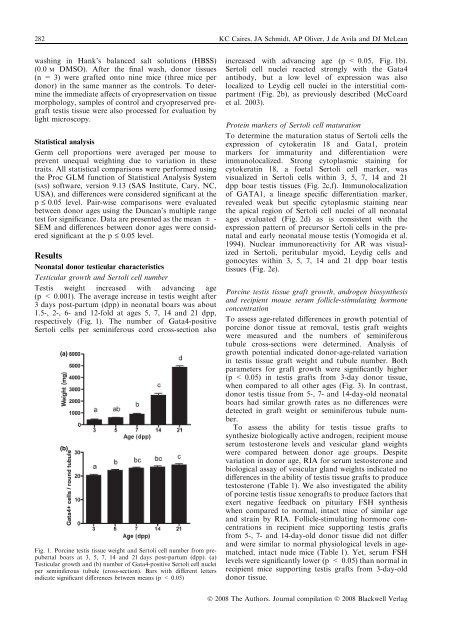
282 KC Caires, JA Schmidt, AP Oliver, J de Avila and DJ McLeanwash<strong>in</strong>g <strong>in</strong> Hank’s balanced salt solutions (HBSS)(0.0 M DMSO). After the f<strong>in</strong>al wash, donor tissues(n = 3) were grafted onto n<strong>in</strong>e mice (three mice perdonor) <strong>in</strong> the same manner as the controls. To determ<strong>in</strong>ethe immediate affects of cryopreservation on tissuemorphology, samples of control and cryopreserved pregrafttestis tissue were also processed for evaluation bylight microscopy.Statistical analysisGerm cell proportions were averaged per mouse toprevent unequal weight<strong>in</strong>g due to variation <strong>in</strong> thesetraits. All statistical comparisons were performed us<strong>in</strong>gthe Proc GLM function of Statistical Analysis System(SAS) software, version 9.13 (SAS Institute, Cary, NC,USA), and differences were considered significant at thep £ 0.05 level. Pair-wise comparisons were evaluatedbetween donor ages us<strong>in</strong>g the Duncan’s multiple rangetest for significance. Data are presented as the mean ± -SEM and differences between donor ages were consideredsignificant at the p £ 0.05 level.ResultsNeonatal donor testicular characteristicsTesticular growth and Sertoli cell numberTestis weight <strong>in</strong>creased with advanc<strong>in</strong>g age(p < 0.001). The average <strong>in</strong>crease <strong>in</strong> testis weight after3 days post-partum (dpp) <strong>in</strong> neonatal boars was about1.5-, 2-, 6- and 12-fold at ages 5, 7, 14 and 21 dpp,respectively (Fig. 1). The number of Gata4-positiveSertoli cells per sem<strong>in</strong>iferous cord cross-section also(a)(b)Fig. 1. Porc<strong>in</strong>e testis tissue weight and Sertoli cell number from prepubertalboars at 3, 5, 7, 14 and 21 days post-partum (dpp). (a)Testicular growth and (b) number of Gata4-positive Sertoli cell nucleiper sem<strong>in</strong>iferous tubule (cross-section). Bars with different letters<strong>in</strong>dicate significant differences between means (p < 0.05)<strong>in</strong>creased with advanc<strong>in</strong>g age (p < 0.05, Fig. 1b).Sertoli cell nuclei reacted strongly with the Gata4antibody, but a low level of expression was alsolocalized to Leydig cell nuclei <strong>in</strong> the <strong>in</strong>terstitial compartment(Fig. 2b), as previously described (McCoardet al. 2003).Prote<strong>in</strong> markers of Sertoli cell maturationTo determ<strong>in</strong>e the maturation status of Sertoli cells theexpression of cytokerat<strong>in</strong> 18 and Gata1, prote<strong>in</strong>markers for immaturity and differentiation wereimmunolocalized. Strong cytoplasmic sta<strong>in</strong><strong>in</strong>g forcytokerat<strong>in</strong> 18, a foetal Sertoli cell marker, wasvisualized <strong>in</strong> Sertoli cells with<strong>in</strong> 3, 5, 7, 14 and 21dpp boar testis tissues (Fig. 2c,f). Immunolocalizationof GATA1, a l<strong>in</strong>eage specific differentiation marker,revealed weak but specific cytoplasmic sta<strong>in</strong><strong>in</strong>g nearthe apical region of Sertoli cell nuclei of all neonatalages evaluated (Fig. 2d) as is consistent with theexpression pattern of precursor Sertoli cells <strong>in</strong> the prenataland early neonatal mouse testis (Yomogida et al.1994). Nuclear immunoreactivity for AR was visualized<strong>in</strong> Sertoli, peritubular myoid, Leydig cells andgonocytes with<strong>in</strong> 3, 5, 7, 14 and 21 dpp boar testistissues (Fig. 2e).Porc<strong>in</strong>e testis tissue graft growth, androgen biosynthesisand recipient mouse serum follicle-stimulat<strong>in</strong>g hormoneconcentrationTo assess age-related differences <strong>in</strong> growth potential ofporc<strong>in</strong>e donor tissue at removal, testis graft weightswere measured and the numbers of sem<strong>in</strong>iferoustubule cross-sections were determ<strong>in</strong>ed. Analysis ofgrowth potential <strong>in</strong>dicated donor-age-related variation<strong>in</strong> testis tissue graft weight and tubule number. Bothparameters for graft growth were significantly higher(p < 0.05) <strong>in</strong> testis grafts from 3-day donor tissue,when compared to all other ages (Fig. 3). In contrast,donor testis tissue from 5-, 7- and 14-day-old neonatalboars had similar growth rates as no differences weredetected <strong>in</strong> graft weight or sem<strong>in</strong>iferous tubule number.To assess the ability for testis tissue grafts tosynthesize biologically active androgen, recipient mouseserum testosterone levels and vesicular gland weightswere compared between donor age groups. Despitevariation <strong>in</strong> donor age, RIA for serum testosterone andbiological assay of vesicular gland weights <strong>in</strong>dicated nodifferences <strong>in</strong> the ability of testis tissue grafts to producetestosterone (Table 1). We also <strong>in</strong>vestigated the abilityof porc<strong>in</strong>e testis tissue xenografts to produce factors thatexert negative feedback on pituitary FSH synthesiswhen compared to normal, <strong>in</strong>tact mice of similar ageand stra<strong>in</strong> by RIA. Follicle-stimulat<strong>in</strong>g hormone concentrations<strong>in</strong> recipient mice support<strong>in</strong>g testis graftsfrom 5-, 7- and 14-day-old donor tissue did not differand were similar to normal physiological levels <strong>in</strong> agematched,<strong>in</strong>tact nude mice (Table 1). Yet, serum FSHlevels were significantly lower (p < 0.05) than normal <strong>in</strong>recipient mice support<strong>in</strong>g testis grafts from 3-day-olddonor tissue.Ó 2008 The Authors. Journal compilation Ó 2008 Blackwell Verlag


