Reproduction in Domestic Animals
Reproduction in Domestic Animals
Reproduction in Domestic Animals
- No tags were found...
You also want an ePaper? Increase the reach of your titles
YUMPU automatically turns print PDFs into web optimized ePapers that Google loves.
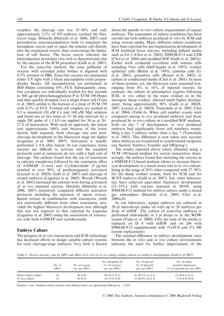
334 C Galli, I Lagut<strong>in</strong>a, R Duchi, S Colleoni and G Lazzaricouplets, the cleavage rate was 35–50% and onlyapproximately 2.5% of NT-embryos reached the blastocyststage. H<strong>in</strong>richs (H<strong>in</strong>richs et al. 2006, 2007) usedpiezoelectric micromanipulation both to enucleate themetaphase oocyte and to <strong>in</strong>ject the somatic cell directly<strong>in</strong>to the enucleated oocyte, thus overcom<strong>in</strong>g the limitationof cell fusion. The horse oocyte tolerates themicro<strong>in</strong>jection procedure very well as demonstrated alsoby the success of the ICSI procedure (Galli et al. 2007).For the zona-free method, the zona pellucida ofoocytes with extruded polar bodies is digested with0.5% pronase <strong>in</strong> PBS. Zona-free oocytes are enucleatedunder UV light with a blunt micropipette (with perpendicularbreak). All manipulations are performed <strong>in</strong>SOF-Hepes conta<strong>in</strong><strong>in</strong>g 10% FCS. Subsequently, zonafreecytoplasts are <strong>in</strong>dividually washed for few seconds<strong>in</strong> 300 lg ⁄ ml phytohemagglut<strong>in</strong><strong>in</strong> P <strong>in</strong> TCM 199-Hepesand then quickly dropped over a s<strong>in</strong>gle donor cell (Vajtaet al. 2003) settled to the bottom of a drop of TCM 199with 0.5% of FCS. Formed cell couplets are washed <strong>in</strong>0.3 M mannitol (50 lM Ca and 100 lM Mg) solutionand fused one or two times at 15–30 m<strong>in</strong> <strong>in</strong>tervals by as<strong>in</strong>gle DC-pulse of 1.2 kV ⁄ cm applied for 30 ls at 26–27 h of maturation. With such an approach, the fusionrate approximates 100% and because of the lowerelectric field required, both cleavage rate and postcleavagedevelopment to the blastocyst stage are higher(Lagut<strong>in</strong>a et al. 2005, 2007). Activation is usuallyperformed 2–4 h after fusion. In our experience, horseoocytes are difficult to activate and the standardprotocols used <strong>in</strong> rum<strong>in</strong>ants do not yield a high rate ofcleavage. The authors found that the use of ionomyc<strong>in</strong>(a calcium ionophore) followed by the synergistic effectof 6-DMAP (1 mM) and Cycloheximide (5 lg ⁄ ml)resulted <strong>in</strong> over 90% of parthenogenetic activation(Lazzari et al. 2002b; Galli et al. 2007) and cleavage ofcloned embryos (Lagut<strong>in</strong>a et al. 2005). Woods (Woodset al. 2003) <strong>in</strong>creased the calcium level dur<strong>in</strong>g activationof <strong>in</strong> vivo matured oocytes. H<strong>in</strong>richs (H<strong>in</strong>richs et al.2006, 2007) extensively compared different activationprotocols <strong>in</strong>clud<strong>in</strong>g the <strong>in</strong>jection of sperm extract.Sperm extract <strong>in</strong> comb<strong>in</strong>ation with ionomyc<strong>in</strong>, whilenot statistically different from other treatments, providedthe highest blastocyst development rate althoughthis was not superior to that reported by Lagut<strong>in</strong>a(Lagut<strong>in</strong>a et al. 2005) us<strong>in</strong>g the association of ionomyc<strong>in</strong>with both 6-DMAP and cycloheximide.Embryo CultureThe progress <strong>in</strong> <strong>in</strong> vitro maturation and ICSI technologyhas <strong>in</strong>creased efforts to design suitable culture systemsfor early cleavage-stage embryos. Very little is knownabout the specific <strong>in</strong> vitro culture requirements of equ<strong>in</strong>eembryos. The assessment of culture conditions has beencarried out with embryos produced <strong>in</strong> vitro by ICSI andus<strong>in</strong>g various media. Many different culture conditionshave been reported for pre-implantation development ofICSI fertilized horse oocytes, <strong>in</strong>clud<strong>in</strong>g def<strong>in</strong>ed mediasuch as G1.2 (Choi et al. 2002), DMEM-F12 and CZB(Choi et al. 2004) and modified SOF (Galli et al. 2002a).Earlier work evaluated co-culture with somatic cells<strong>in</strong>clud<strong>in</strong>g Vero cells (Dell’Aquila et al. 1997), oviductepithelial cells (Battut et al. 1991), cumulus cells (Liet al. 2001), granulosa cells (Rosati et al. 2002) orculture <strong>in</strong> conditioned media (Choi et al. 2001). In mostof these systems, yet, the blastocyst rates rema<strong>in</strong>ed low,rang<strong>in</strong>g from 4% to 16% of <strong>in</strong>jected oocytes. Incontrast, the culture of presumptive zygotes follow<strong>in</strong>gICSI, <strong>in</strong> vivo either <strong>in</strong> the mare oviduct or <strong>in</strong> thesurrogate sheep oviduct, allowed much higher development,be<strong>in</strong>g approximately 30% (Galli et al. 2002b,2007; Lazzari et al. 2002b; Tremoleda et al. 2003; Choiet al. 2004) (Table 6). When cell number counts werecompared among <strong>in</strong> vivo produced embryos and thoseproduced by <strong>in</strong> vitro culture <strong>in</strong> a modified SOF medium,both on day 7 of development, <strong>in</strong> vitro producedembryos had significantly lower cell numbers, resembl<strong>in</strong>ga day 5 embryo rather than a day 7 (Tremoledaet al. 2003). This difference is now taken <strong>in</strong>to accountwhen embryos are transferred to synchronized recipients(see Section ‘Embryo Transfer and Offspr<strong>in</strong>g’).The results reported above where obta<strong>in</strong>ed us<strong>in</strong>g aTCM 199-based medium for oocyte maturation. Interest<strong>in</strong>gly,the authors found that matur<strong>in</strong>g the oocytes <strong>in</strong>a DMEM-F12-based medium allows to <strong>in</strong>crease blastocystdevelopment to a much closer rate to <strong>in</strong> vivo culture,be<strong>in</strong>g <strong>in</strong> the range of 26% when compared with 27–50%for the sheep oviduct system, both for ICSI and forSCNT embryos (Galli et al. 2007). Yet, other laboratorieshave achieved equivalent blastocyst development(25–35%) with oocytes matured <strong>in</strong> M199, us<strong>in</strong>gDMEM-F12 medium for embryo culture under a mixedgas atmosphere (H<strong>in</strong>richs et al. 2005; Choi et al.2006a,b).In our laboratory, equ<strong>in</strong>e embryos are cultured <strong>in</strong>20 ll microdrops under oil with up to 20 embryos perdrop of mSOF. The culture of zona-free embryos isperformed <strong>in</strong>dividually <strong>in</strong> 5 ll drops or <strong>in</strong> the WOWsystem (Vajta et al. 2000). Fifty per cent of the media isreplaced on D 4 with mSOF and on D6 withDMEM ⁄ F12 supplemented with 5%FCS and 5% SR(serum replacement).The residual difference <strong>in</strong> embryo development ratesbetween the <strong>in</strong> vitro and <strong>in</strong> vivo culture environments<strong>in</strong>dicates the need for further improvement of theTable 6. Oocyte recovery rate by OPU and effect of <strong>in</strong> vitro or <strong>in</strong> vivo sheep oviduct culture on embryos development (Galli et al 2007)No. ofOPUsNo. of oocytes(no. per OPU)No. metaphase II(% of oocytes)(no. per OPU)No. of cleaved(% of <strong>in</strong>jected)(no. per OPU)No. of comp.morulae ⁄ blastocysts(% of <strong>in</strong>jected) (no. per OPU)Sheep oviduct culture 20 60 (3.0) 46 (76.7) (2.3) 41 (89.1% a) (2.1) 23 (50.0 a) (1.2)In vitro culture 12 46 (4.1) 36 (73.5) (3.0) 25 (69.4% a) (2.1) 5 (13.9 b) (0.4)Student’s t-test. Numbers with<strong>in</strong> columns with different letters are significantly different (p < 0.05).Ó 2008 The Authors. Journal compilation Ó 2008 Blackwell Verlag


