

Reproduction in Domestic Animals
Reproduction in Domestic Animals
Reproduction in Domestic Animals
- No tags were found...

Create successful ePaper yourself
Turn your PDF publications into a flip-book with our unique Google optimized e-Paper software.
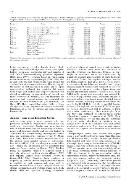
326 CR Barb, GJ Hausman and CA LentsTable 1. Microarray analysis of several fat depots demonstrated that thyroid hormone receptors were collectively upregulated while several otherreceptors (MCIR, FGF4) and lipogenic (LPL, SCD) enzymes were downregulated with fast<strong>in</strong>g aFat depot Gene Estimate SE T-value p-valuePerirenal Thyroid hormone receptor alpha 2 (Sus scrofa) )0.20083 0.051199 )3.92248 0.000254Mesenteric Thyroid hormone receptor, alpha )0.16131 0.047101 )3.42473 0.001195Leaf Thyroid hormone receptor, alpha )0.15323 0.030834 )4.96954 0.000007Leaf Thyroid hormone receptor alpha 2 (Sus scrofa) )0.31586 0.089853 )3.51524 0.000909Leaf Melanocort<strong>in</strong> 1 receptor 0.357846 0.175065 2.044075 0.045929Perirenal Melanocort<strong>in</strong> 1 receptor 0.396369 0.117805 3.364614 0.001431Leaf Fibroblast growth factor receptor 4 0.40214 0.121846 3.300404 0.001731Perirenal Fibroblast growth factor receptor 4 0.44749 0.139023 3.218823 0.002199Perirenal Stearoyl-CoA desaturase 0.914822 0.323623 2.826815 0.006618Lipoprote<strong>in</strong> lipase 0.43321 0.125486 3.452266 0.0011Mesenteric Stearoyl-CoA desaturase 1.40823 0.304109 4.630675 0.000024Lipoprote<strong>in</strong> lipase 0.14577 0.069703 2.091302 0.04131Leaf Stearoyl-CoA desaturase 0.170878 0.071794 2.380111 0.020937Lipoprote<strong>in</strong> lipase 0.380183 0.079236 4.798133 0.000013a Analysis of the microarray data was conducted with either LOWESS normalization based or an ANOVA normalization based method to determ<strong>in</strong>e the <strong>in</strong>fluence offast<strong>in</strong>g on gene expression.lept<strong>in</strong> secretion or to affect backfat lept<strong>in</strong>, Ob-rb,adipocyte fatty acid b<strong>in</strong>d<strong>in</strong>g prote<strong>in</strong>, or the transcriptionfactors peroxisome proliferator-activated receptor-c2and CCAAT-enhancer-b<strong>in</strong>d<strong>in</strong>g prote<strong>in</strong>-a expression(Hart et al. 2007). However, based on ma<strong>in</strong>tenancerequirements for the pre-pubertal gilt (NRC 1998) used<strong>in</strong> that study, the feed restricted gilts were actually fed124% of the ma<strong>in</strong>tenance requirement. This may expla<strong>in</strong>the failure of feed restriction to affect LH or lept<strong>in</strong>concentrations. Although feed restriction did preventbody weight and backfat ga<strong>in</strong> adipocyte function wasaltered as evidenced by upregulation of thyroid hormonereceptor-a <strong>in</strong> perirenal, leaf and mesenteric fatdepots <strong>in</strong> feed restricted animals; this co<strong>in</strong>cided withelevated thyrox<strong>in</strong> concentration (GJ Hausman, CRBarb, HA Hart, unpublished data; Table 1). Thesechanges may <strong>in</strong> part represent an attempt to ma<strong>in</strong>ta<strong>in</strong>thermogenesis, as well as immune and neruoendocr<strong>in</strong>ehomeostasis.Adipose Tissue as an Endocr<strong>in</strong>e OrganAdipose tissue plays a more dynamic role thanpreviously thought <strong>in</strong> physiological mechanisms andwhole-body homeostasis. Studies now show that therole of adipose tissue <strong>in</strong>cludes respond<strong>in</strong>g to nutrient,neural and hormonal signals, and secret<strong>in</strong>g factors or‘adipok<strong>in</strong>es’ that control feed<strong>in</strong>g, thermogenesis, immunity,and neuroendocr<strong>in</strong>e function (review by Ahimaet al. 2006). The current evidence <strong>in</strong>dicates that of allthe adipose tissue secreted factors, or ‘adipok<strong>in</strong>es’,<strong>in</strong>terleuk<strong>in</strong>s (IL)-6, IL-8, plasm<strong>in</strong>ogen activator <strong>in</strong>hibitor-1,lept<strong>in</strong> and adiponect<strong>in</strong> can be considered trueendocr<strong>in</strong>e factors (review by Hauner 2005). Furthermore,the secretory function of adipose tissue isadversely <strong>in</strong>fluenced by both obesity (Hauner 2005)and impaired adipose tissue accretion (review byGuerre-Millo 2004). Therefore, body condition oradiposity by virtue of secreted adipok<strong>in</strong>es could impactor regulate, <strong>in</strong> part, physiological states such asreproductive condition or status. Adipose tissue constitutesthe largest amount of stored energy <strong>in</strong> the body(Loftus 1999), and regulation of energy expenditure<strong>in</strong>volves a balance of several factors, such as feed<strong>in</strong>gbehaviour, adipose tissue mass and activation ofcatabolic processes (e.g. lactation). Changes <strong>in</strong> bodyweight or nutritional status are characterized byalterations <strong>in</strong> serum concentrations of many hormonesand growth factors that regulate adipocyte functionand lept<strong>in</strong> secretion (Barb et al. 2001b). Recent microarrayanalyses by our laboratory revealed that 21 genesencod<strong>in</strong>g secreted prote<strong>in</strong>s were expressed 40-fold overbackground <strong>in</strong> neonatal porc<strong>in</strong>e adipose tissue andporc<strong>in</strong>e pre-adipocyte cultures (Hausman et al. 2006).Additionally, agouti gene expression was detected byRT-PCR <strong>in</strong> pig adipose tissue. Proteomic analysis ofadipocyte culture conditioned media identified severalsecreted prote<strong>in</strong>s, <strong>in</strong>clud<strong>in</strong>g several neurotrophic factors,IL-1A, IL-1B, IL-8, IL-6, IL-15, and IGF b<strong>in</strong>d<strong>in</strong>gprote<strong>in</strong>-5. Through microarray and RT-PCR analyses,we recently demonstrated that <strong>in</strong> addition to these,several other cytok<strong>in</strong>es, e.g., ciliary neurotrophic factorand NPY, are expressed by adipose tissue dur<strong>in</strong>gpubertal development (Hausman et al. 2007). Thesestudies demonstrate for the first time the expressionof several major adipok<strong>in</strong>es or cytok<strong>in</strong>es <strong>in</strong> pigadipose tissue which may <strong>in</strong>fluence local and centralmetabolism and growth. Thus, these reports supportthe idea that adipose tissue functions as an endocr<strong>in</strong>eorgan.Morphological studies have revealed that adiposetissue is <strong>in</strong>nervated by adrenergic nerve fibres (Hausmanand Richardson 1987). Further, immunocytochemicaldata revealed that most of the subpopulations of theadrenergic lept<strong>in</strong> receptor immuno-reactive (OBR-IR)neurones supply<strong>in</strong>g fat tissue <strong>in</strong> the pig were positive forNPY and tyros<strong>in</strong>e hydroxylase immunoactivity (Czajaet al. 2002). Moreover, immuno-positive neurones forOBR were located <strong>in</strong> the paraventricular nucleus,ventromedial nucleus, anterior hypothalamic area, preopticarea, arcuate nucleus and supraoptic nucleus(Czaja et al. 2003). Collectively, these studies providemorphological data demonstrat<strong>in</strong>g that hypothalamicOBR conta<strong>in</strong><strong>in</strong>g neurones are transsynaptically connectedto the perirenal fat depot. Therefore, the aboveevidence supports a direct l<strong>in</strong>k between hypothalamicÓ 2008 No claim to orig<strong>in</strong>al government works


