

Reproduction in Domestic Animals
Reproduction in Domestic Animals
Reproduction in Domestic Animals
- No tags were found...

Create successful ePaper yourself
Turn your PDF publications into a flip-book with our unique Google optimized e-Paper software.
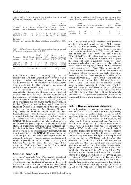
Clon<strong>in</strong>g <strong>in</strong> Horses 333Table 3. Effect of maturation media on maturation, cleavage rate andICSI embryo development (Galli et al. 2007)MaturationmediumNo. of No. ofoocytes degeneratedNo. ofmetaphaseII (%)No. of<strong>in</strong>jectedNo. ofcleaved(% of<strong>in</strong>jected)No. ofblastocysts(% of<strong>in</strong>jected)TCM 199 434 105 205 (47.2 a) 191 111 (58.1 a) 23 (12.0 a)DMEM ⁄ F12 338 71 159 (45.6 a) 140 108 (77.1 b) 37 (26.4 b)Replicates = 6.Chi square test. Numbers with<strong>in</strong> columns with different letters differ (p < 0.05).Table 4. Effect of maturation media on maturation, cleavage rate andNT embryo development (Galli et al. 2007)MaturationmediumNo. ofoocytesNo. oflysedNo. ofmetaphaseII (%)No. ofNTembryosNo. ofcleaved(% of NT)No. ofblastocysts(% of NT)TCM 199 164 35 64 (39.0 a) 41 38 (92.7 a) 4 (9.77 a)DMEM ⁄ F12 166 35 67 (40.4 a) 47 46 (97.9 a) 13 (27.7 b)Chi square test. Numbers with<strong>in</strong> columns with different letters differ (p < 0.05).(H<strong>in</strong>richs et al. 2005). In that study, high rates ofdegeneration <strong>in</strong> culture were seen only <strong>in</strong> oocytes with acompact cumulus; evaluation of oocyte chromat<strong>in</strong>configurations led to the conclusion that these oocyteswere juvenile and that their chromat<strong>in</strong> was damageddur<strong>in</strong>g storage with<strong>in</strong> the ovary.It is known that <strong>in</strong> vitro maturation conditionsdramatically <strong>in</strong>fluence the development of fertilizedoocytes to the blastocyst stage. Different media are usedfor maturation of equ<strong>in</strong>e oocytes, although the mostcommon rema<strong>in</strong>s medium TCM199, probably becauseof its widespread use for bov<strong>in</strong>e oocyte maturation. Inthe last 2 years, the authors have tested other media<strong>in</strong>clud<strong>in</strong>g DMEM-F12 supplemented with 15% serumreplacement (Invitrogen sri, San Giuliano Milanese,Italy) <strong>in</strong>stead of TCM 199 supplemented with 10%foetal calf serum (FCS). Hormones and growth factorswere added to both media as reported earlier (Lagut<strong>in</strong>aet al. 2005). We found a clear advantage <strong>in</strong> the use of aDMEM-F12 based medium not only on cleavage ratebut also on embryo development to the blastocyst stage(Table 3). Prelim<strong>in</strong>ary experiments have confirmed similarf<strong>in</strong>d<strong>in</strong>gs <strong>in</strong> horse SCNT (Table 4). This positiveeffect is evident also on blastocyst development, which issignificantly higher <strong>in</strong> the DMEM-F12 group than <strong>in</strong> theTCM199 group. This remarkable improvement <strong>in</strong>creasesthe efficiency of equ<strong>in</strong>e embryo production andmakes it possible to use a totally <strong>in</strong> vitro systemfollow<strong>in</strong>g OPU <strong>in</strong> the horse. The time required forcomplet<strong>in</strong>g IVM ranges from 22 to 24 h for expandedoocytes and 26–36 h for compacted onesSource of Donor CellsA variety of donor cells from different orig<strong>in</strong> (foetal andadult) and various tissues have been used for SCNTwithout observ<strong>in</strong>g significant differences <strong>in</strong> the overallefficiency (Oback and Wells 2002). In the horse, foetalfibroblasts (Li et al. 2003; Lagut<strong>in</strong>a et al. 2005; WoodsTable 5. Cleavage and blastocyst development after nuclear transferwith confluent or roscovit<strong>in</strong>e-treated fibroblasts (H<strong>in</strong>richs et al. 2006)TreatmentNo. of<strong>in</strong>jected(MII)No. of Cleavage No. of D +cleaved rate 7 blastocystsNo. of D +8 blastocystsTotalblastocyst(%)Confluent 55 40 73 1 1 2 (3.6)Roscovit<strong>in</strong>e 56 44 79 0 2 2 (3.6)et al. 2003) as well as adult fibroblasts and granulosacells have been used (Vanderwall et al. 2004; Lagut<strong>in</strong>aet al. 2005). For recover<strong>in</strong>g adult fibroblasts, sk<strong>in</strong>biopsies are taken under local anaesthesia on the neckor the chest of the donor horse. The recovered tissue isthen m<strong>in</strong>ced <strong>in</strong>to small pieces that are plated <strong>in</strong>TCM199 ⁄ DMEM (1:1) culture medium supplementedwith 10% FCS. In 2–3 weeks, the fibroblasts outgrowthe tissue and form a confluent monolayer. Uponsubsequent sub-culture and expansion, the cells arefrozen for later use or prepared for the SCNT procedureat early passages (Li et al. 2003). There is a considerablevariation <strong>in</strong> the success rate of horse SCNT because ofthe specific cell l<strong>in</strong>e source of donor nuclei (Galli et al.2003a; Lagut<strong>in</strong>a et al. 2005) as reported for other species(Oback and Wells 2002). The cell cycle of the donor cellis crucial for success and G0 or G1 stages have beenused <strong>in</strong> horses. This stages can be achieved throughserum starvation (reduction of FCS to 0.5%), growth toconfluency (contact <strong>in</strong>hibition) or the use of k<strong>in</strong>ase<strong>in</strong>hibitors like Roscovit<strong>in</strong>e (Table 5) (Oback and Wells2002; H<strong>in</strong>richs et al. 2006, 2007). Given the relativelylow number of experiments performed, it cannot beascerta<strong>in</strong>ed if any of these approaches is better than theother.Embryo Reconstruction and ActivationIn our laboratory, the oocytes are stripped of theircumulus after 22–24 h of maturation. First, by pipett<strong>in</strong>gdur<strong>in</strong>g exposure to hyaluronidase; second, <strong>in</strong> 0.25%tryps<strong>in</strong> for 1.5 m<strong>in</strong> and f<strong>in</strong>ally, <strong>in</strong> SOF-Hepes conta<strong>in</strong><strong>in</strong>g10% FCS. For reconstruction of NT-embryos, theauthors use both zona-enclosed and zona-free methods(Oback et al. 2003). For the zona-enclosed method,oocytes with an extruded polar body are sta<strong>in</strong>ed withHoechst 33342 <strong>in</strong> the presence of cytochalas<strong>in</strong> B(5 lg ⁄ ml). Enucleation is performed by the aspirationof the polar body and metaphase II plate <strong>in</strong> a m<strong>in</strong>imalvolume of ooplasm under UV light. All manipulationsare carried out <strong>in</strong> SOF-Hepes with 6 mg ⁄ ml BSA(Gardner et al. 1994), except fusion. Individual nucleardonor cells are then transferred <strong>in</strong>to the perivitell<strong>in</strong>espace of zona-enclosed cytoplasts us<strong>in</strong>g the enucleationpipette. The cytoplast–karyoplast couplets are transferred<strong>in</strong>to 0.3 M mannitol (50 lM Ca and 100 lM Mg)solution and fused one or two times with<strong>in</strong> 15–30 m<strong>in</strong><strong>in</strong>terval by a double DC-pulse of 1.8–2.4 kV ⁄ cm appliedfor 30 ls at 26–27 h of maturation. Because of theheterogeneous characteristics of the horse zona pellucida,the fusion rate is often <strong>in</strong>consistent or low. Li et al.(2002) have found that even high voltage DC pulses of2.2–2.5 kV ⁄ cm were able to fuse less than 60% ofÓ 2008 The Authors. Journal compilation Ó 2008 Blackwell Verlag


