

Reproduction in Domestic Animals
Reproduction in Domestic Animals
Reproduction in Domestic Animals
- No tags were found...

You also want an ePaper? Increase the reach of your titles
YUMPU automatically turns print PDFs into web optimized ePapers that Google loves.
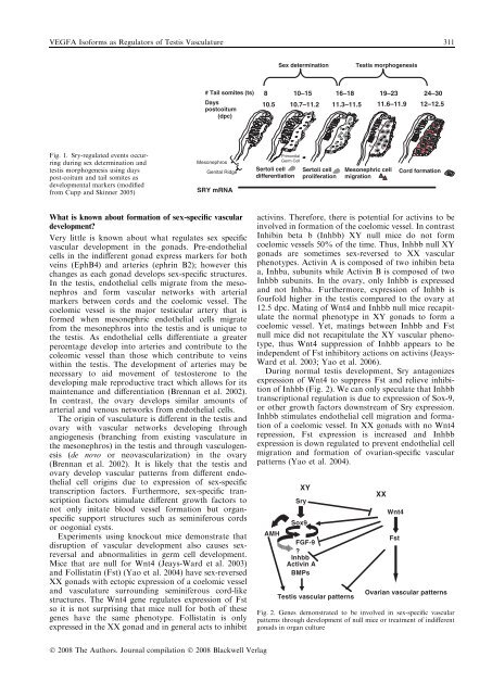
VEGFA Isoforms as Regulators of Testis Vasculature 311Sex determ<strong>in</strong>ationTestis morphogenesis# Tail somites (ts)Da yspostcoitum(dpc)8 10–15 16–18 19–23 24–3010. 5 10.7–11.2 11.3–11.5 11.6–11.9 12–12.5Fig. 1. Sry-regulated events occurr<strong>in</strong>gdur<strong>in</strong>g sex determ<strong>in</strong>ation andtestis morphogenesis us<strong>in</strong>g dayspost-coitum and tail somites asdevelopmental markers (modifiedfrom Cupp and Sk<strong>in</strong>ner 2005)MesonephrosGenital RidgeSRY mRN APrim ordialGe rm CellSertoli celldifferentiationSertoli cellproliferationMesonephric ce llmigrationCord formatio nWhat is known about formation of sex-specific vasculardevelopment?Very little is known about what regulates sex specificvascular development <strong>in</strong> the gonads. Pre-endothelialcells <strong>in</strong> the <strong>in</strong>different gonad express markers for bothve<strong>in</strong>s (EphB4) and arteries (ephr<strong>in</strong> B2); however thischanges as each gonad develops sex-specific structures.In the testis, endothelial cells migrate from the mesonephrosand form vascular networks with arterialmarkers between cords and the coelomic vessel. Thecoelomic vessel is the major testicular artery that isformed when mesonephric endothelial cells migratefrom the mesonephros <strong>in</strong>to the testis and is unique tothe testis. As endothelial cells differentiate a greaterpercentage develop <strong>in</strong>to arteries and contribute to thecoleomic vessel than those which contribute to ve<strong>in</strong>swith<strong>in</strong> the testis. The development of arteries may benecessary to aid movement of testosterone to thedevelop<strong>in</strong>g male reproductive tract which allows for itsma<strong>in</strong>tenance and differentiation (Brennan et al. 2002).In contrast, the ovary develops similar amounts ofarterial and venous networks from endothelial cells.The orig<strong>in</strong> of vasculature is different <strong>in</strong> the testis andovary with vascular networks develop<strong>in</strong>g throughangiogenesis (branch<strong>in</strong>g from exist<strong>in</strong>g vasculature <strong>in</strong>the mesonephros) <strong>in</strong> the testis and through vasculogenesis(de novo or neovascularization) <strong>in</strong> the ovary(Brennan et al. 2002). It is likely that the testis andovary develop vascular patterns from different endothelialcell orig<strong>in</strong>s due to expression of sex-specifictranscription factors. Furthermore, sex-specific transcriptionfactors stimulate different growth factors tonot only <strong>in</strong>itate blood vessel formation but organspecificsupport structures such as sem<strong>in</strong>iferous cordsor oogonial cysts.Experiments us<strong>in</strong>g knockout mice demonstrate thatdisruption of vascular development also causes sexreversaland abnormalities <strong>in</strong> germ cell development.Mice that are null for Wnt4 (Jeays-Ward et al. 2003)and Follistat<strong>in</strong> (Fst) (Yao et al. 2004) have sex-reversedXX gonads with ectopic expression of a coelomic vesseland vasculature surround<strong>in</strong>g sem<strong>in</strong>iferous cord-likestructures. The Wnt4 gene regulates expression of Fstso it is not surpris<strong>in</strong>g that mice null for both of thesegenes have the same phenotype. Follistat<strong>in</strong> is onlyexpressed <strong>in</strong> the XX gonad and <strong>in</strong> general acts to <strong>in</strong>hibitactiv<strong>in</strong>s. Therefore, there is potential for activ<strong>in</strong>s to be<strong>in</strong>volved <strong>in</strong> formation of the coelomic vessel. In contrastInhib<strong>in</strong> beta b (Inhbb) XY null mice do not formcoelomic vessels 50% of the time. Thus, Inhbb null XYgonads are sometimes sex-reversed to XX vascularphenotypes. Activ<strong>in</strong> A is composed of two <strong>in</strong>hib<strong>in</strong> betaa, Inhba, subunits while Activ<strong>in</strong> B is composed of twoInhbb subunits. In the ovary, only Inhbb is expressedand not Inhba. Furthermore, expression of Inhbb isfourfold higher <strong>in</strong> the testis compared to the ovary at12.5 dpc. Mat<strong>in</strong>g of Wnt4 and Inhbb null mice recapitulatethe normal phenotype <strong>in</strong> XY gonads to form acoelomic vessel. Yet, mat<strong>in</strong>gs between Inhbb and Fstnull mice did not recapitulate the XY vascular phenotype,thus Wnt4 suppression of Inhbb appears to be<strong>in</strong>dependent of Fst <strong>in</strong>hibitory actions on activ<strong>in</strong>s (Jeays-Ward et al. 2003; Yao et al. 2006).Dur<strong>in</strong>g normal testis development, Sry antagonizesexpression of Wnt4 to suppress Fst and relieve <strong>in</strong>hibitionof Inhbb (Fig. 2). We can only speculate that Inhbbtranscriptional regulation is due to expression of Sox-9,or other growth factors downstream of Sry expression.Inhbb stimulates endothelial cell migration and formationof a coelomic vessel. In XX gonads with no Wnt4repression, Fst expression is <strong>in</strong>creased and Inhbbexpression is down regulated to prevent endothelial cellmigration and formation of ovarian-specific vascularpatterns (Yao et al. 2004).AMHXYSrySox9FGF-9?InhbbActiv<strong>in</strong> ABMPsTestis vascular patternsXXWnt4FstOvarian vascular patternsFig. 2. Genes demonstrated to be <strong>in</strong>volved <strong>in</strong> sex-specific vascularpatterns through development of null mice or treatment of <strong>in</strong>differentgonads <strong>in</strong> organ cultureÓ 2008 The Authors. Journal compilation Ó 2008 Blackwell Verlag


