

Reproduction in Domestic Animals
Reproduction in Domestic Animals
Reproduction in Domestic Animals
- No tags were found...

Create successful ePaper yourself
Turn your PDF publications into a flip-book with our unique Google optimized e-Paper software.
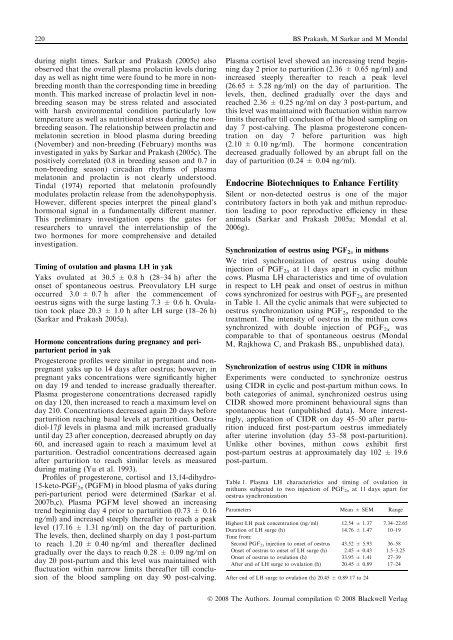
220 BS Prakash, M Sarkar and M Mondaldur<strong>in</strong>g night times. Sarkar and Prakash (2005c) alsoobserved that the overall plasma prolact<strong>in</strong> levels dur<strong>in</strong>gday as well as night time were found to be more <strong>in</strong> nonbreed<strong>in</strong>gmonth than the correspond<strong>in</strong>g time <strong>in</strong> breed<strong>in</strong>gmonth. This marked <strong>in</strong>crease of prolact<strong>in</strong> level <strong>in</strong> nonbreed<strong>in</strong>gseason may be stress related and associatedwith harsh environmental condition particularly lowtemperature as well as nutritional stress dur<strong>in</strong>g the nonbreed<strong>in</strong>gseason. The relationship between prolact<strong>in</strong> andmelaton<strong>in</strong> secretion <strong>in</strong> blood plasma dur<strong>in</strong>g breed<strong>in</strong>g(November) and non-breed<strong>in</strong>g (February) months was<strong>in</strong>vestigated <strong>in</strong> yaks by Sarkar and Prakash (2005c). Thepositively correlated (0.8 <strong>in</strong> breed<strong>in</strong>g season and 0.7 <strong>in</strong>non-breed<strong>in</strong>g season) circadian rhythms of plasmamelaton<strong>in</strong> and prolact<strong>in</strong> is not clearly understood.T<strong>in</strong>dal (1974) reported that melaton<strong>in</strong> profoundlymodulates prolact<strong>in</strong> release from the adenohypophysis.However, different species <strong>in</strong>terpret the p<strong>in</strong>eal gland’shormonal signal <strong>in</strong> a fundamentally different manner.This prelim<strong>in</strong>ary <strong>in</strong>vestigation opens the gates forresearchers to unravel the <strong>in</strong>terrelationship of thetwo hormones for more comprehensive and detailed<strong>in</strong>vestigation.Tim<strong>in</strong>g of ovulation and plasma LH <strong>in</strong> yakYaks ovulated at 30.5 ± 0.8 h (28–34 h) after theonset of spontaneous oestrus. Preovulatory LH surgeoccurred 3.0 ± 0.7 h after the commencement ofoestrus signs with the surge last<strong>in</strong>g 7.3 ± 0.6 h. Ovulationtook place 20.3 ± 1.0 h after LH surge (18–26 h)(Sarkar and Prakash 2005a).Hormone concentrations dur<strong>in</strong>g pregnancy and periparturientperiod <strong>in</strong> yakProgesterone profiles were similar <strong>in</strong> pregnant and nonpregnantyaks up to 14 days after oestrus; however, <strong>in</strong>pregnant yaks concentrations were significantly higheron day 19 and tended to <strong>in</strong>crease gradually thereafter.Plasma progesterone concentrations decreased rapidlyon day 120, then <strong>in</strong>creased to reach a maximum level onday 210. Concentrations decreased aga<strong>in</strong> 20 days beforeparturition reach<strong>in</strong>g basal levels at parturition. Oestradiol-17blevels <strong>in</strong> plasma and milk <strong>in</strong>creased graduallyuntil day 23 after conception, decreased abruptly on day60, and <strong>in</strong>creased aga<strong>in</strong> to reach a maximum level atparturition. Oestradiol concentrations decreased aga<strong>in</strong>after parturition to reach similar levels as measureddur<strong>in</strong>g mat<strong>in</strong>g (Yu et al. 1993).Profiles of progesterone, cortisol and 13,14-dihydro-15-keto-PGF 2a (PGFM) <strong>in</strong> blood plasma of yaks dur<strong>in</strong>gperi-parturient period were determ<strong>in</strong>ed (Sarkar et al.2007b,c). Plasma PGFM level showed an <strong>in</strong>creas<strong>in</strong>gtrend beg<strong>in</strong>n<strong>in</strong>g day 4 prior to parturition (0.73 ± 0.16ng ⁄ ml) and <strong>in</strong>creased steeply thereafter to reach a peaklevel (17.16 ± 1.31 ng ⁄ ml) on the day of parturition.The levels, then, decl<strong>in</strong>ed sharply on day 1 post-partumto reach 1.20 ± 0.40 ng ⁄ ml and thereafter decl<strong>in</strong>edgradually over the days to reach 0.28 ± 0.09 ng ⁄ ml onday 20 post-partum and this level was ma<strong>in</strong>ta<strong>in</strong>ed withfluctuation with<strong>in</strong> narrow limits thereafter till conclusionof the blood sampl<strong>in</strong>g on day 90 post-calv<strong>in</strong>g.Plasma cortisol level showed an <strong>in</strong>creas<strong>in</strong>g trend beg<strong>in</strong>n<strong>in</strong>gday 2 prior to parturition (2.36 ± 0.65 ng ⁄ ml) and<strong>in</strong>creased steeply thereafter to reach a peak level(26.65 ± 5.28 ng ⁄ ml) on the day of parturition. Thelevels, then, decl<strong>in</strong>ed gradually over the days andreached 2.36 ± 0.25 ng ⁄ ml on day 3 post-partum, andthis level was ma<strong>in</strong>ta<strong>in</strong>ed with fluctuation with<strong>in</strong> narrowlimits thereafter till conclusion of the blood sampl<strong>in</strong>g onday 7 post-calv<strong>in</strong>g. The plasma progesterone concentrationon day 7 before parturition was high(2.10 ± 0.10 ng ⁄ ml). The hormone concentrationdecreased gradually followed by an abrupt fall on theday of parturition (0.24 ± 0.04 ng ⁄ ml).Endocr<strong>in</strong>e Biotechniques to Enhance FertilitySilent or non-detected oestrus is one of the majorcontributory factors <strong>in</strong> both yak and mithun reproductionlead<strong>in</strong>g to poor reproductive efficiency <strong>in</strong> theseanimals (Sarkar and Prakash 2005a; Mondal et al.2006g).Synchronization of oestrus us<strong>in</strong>g PGF 2a <strong>in</strong> mithunsWe tried synchronization of oestrus us<strong>in</strong>g double<strong>in</strong>jection of PGF 2a at 11 days apart <strong>in</strong> cyclic mithuncows. Plasma LH characteristics and time of ovulation<strong>in</strong> respect to LH peak and onset of oestrus <strong>in</strong> mithuncows synchronized for oestrus with PGF 2a are presented<strong>in</strong> Table 1. All the cyclic animals that were subjected tooestrus synchronization us<strong>in</strong>g PGF 2a responded to thetreatment. The <strong>in</strong>tensity of oestrus <strong>in</strong> the mithun cowssynchronized with double <strong>in</strong>jection of PGF 2a wascomparable to that of spontaneous oestrus (MondalM, Rajkhowa C, and Prakash BS., unpublished data).Synchronization of oestrus us<strong>in</strong>g CIDR <strong>in</strong> mithunsExperiments were conducted to synchronize oestrusus<strong>in</strong>g CIDR <strong>in</strong> cyclic and post-partum mithun cows. Inboth categories of animal, synchronized oestrus us<strong>in</strong>gCIDR showed more prom<strong>in</strong>ent behavioural signs thanspontaneous heat (unpublished data). More <strong>in</strong>terest<strong>in</strong>gly,application of CIDR on day 45–50 after parturition<strong>in</strong>duced first post-partum oestrus immediatelyafter uter<strong>in</strong>e <strong>in</strong>volution (day 53–58 post-parturition).Unlike other bov<strong>in</strong>es, mithun cows exhibit firstpost-partum oestrus at approximately day 102 ± 19.6post-partum.Table 1. Plasma LH characteristics and tim<strong>in</strong>g of ovulation <strong>in</strong>mithuns subjected to two <strong>in</strong>jection of PGF 2a at 11 days apart foroestrus synchronizationParameters Mean ± SEM RangeHighest LH peak concentration (ng ⁄ ml) 12.54 ± 1.37 7.34–22.65Duration of LH surge (h) 14.76 ± 1.47 10–19Time from:Second PGF 2a <strong>in</strong>jection to onset of oestrus 43.52 ± 5.93 36–58Onset of oestrus to onset of LH surge (h) 2.45 ± 0.43 1.5–3.25Onset of oestrus to ovulation (h) 33.95 ± 1.41 27–39After end of LH surge to ovulation (h) 20.45 ± 0.89 17–24After end of LH surge to ovulation (h) 20.45 ± 0.89 17 to 24Ó 2008 The Authors. Journal compilation Ó 2008 Blackwell Verlag


