

Reproduction in Domestic Animals
Reproduction in Domestic Animals
Reproduction in Domestic Animals
- No tags were found...

You also want an ePaper? Increase the reach of your titles
YUMPU automatically turns print PDFs into web optimized ePapers that Google loves.
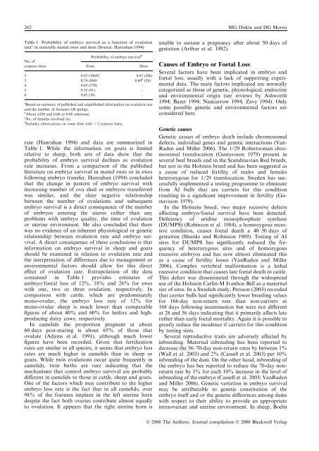
262 MG Disk<strong>in</strong> and DG MorrisTable 1. Probability of embryo survival as a function of ovulationrate a <strong>in</strong> naturally mated ewes and does (Source: Hanrahan 1994)No. ofcorpora luteaEwesProbability of embryo survival bDoes2 0.82 (5069) c 0.82 (306)3 0.74 (884) 0.60 d (58)4 0.65 (270) –5 0.55 (91) –6 0.45 (38) –a Based on summary of published and unpublished <strong>in</strong>formation on ovulation rateand the number of foetuses ⁄ off-spr<strong>in</strong>gs.b About ‡250 and 0.06 to 0.08 otherwise.c No. of females <strong>in</strong>volved (n).d Includes observations on some does with >3 corpora lutea.rate (Hanrahan 1994) and data are summarized <strong>in</strong>Table 1. While the <strong>in</strong>formation on goats is limitedrelative to sheep, both sets of data show that theprobability of embryo survival decl<strong>in</strong>es as ovulationrate <strong>in</strong>creases. From a comparison of the publishedliterature on embryo survival <strong>in</strong> mated ewes or <strong>in</strong> ewesfollow<strong>in</strong>g embryo transfer, Hanrahan (1994) concludedthat the change <strong>in</strong> pattern of embryo survival with<strong>in</strong>creas<strong>in</strong>g number of ova shed or embryos transferredwas similar, and the clear negative relationshipbetween the number of ovulations and subsequentembryo survival is a direct consequence of the numberof embryos enter<strong>in</strong>g the uterus rather than anyproblems with embryo quality, the time of ovulationor uter<strong>in</strong>e environment. He also concluded that therewas no evidence of an <strong>in</strong>herent physiological or geneticrelationship between ovulation rate and embryo survival.A direct consequence of these conclusions is that<strong>in</strong>formation on embryo survival <strong>in</strong> sheep and goatsshould be exam<strong>in</strong>ed <strong>in</strong> relation to ovulation rate andthe <strong>in</strong>terpretation of differences due to management orenvironmental factors should allow for this directeffect of ovulation rate. Extrapolation of the dataconta<strong>in</strong>ed <strong>in</strong> Table 1 provides estimates ofembryo ⁄ foetal loss of 12%, 18% and 26% for eweswith one, two or three ovulation, respectively. Incomparison with cattle, which are predom<strong>in</strong>atelymono-ovular, the embryo loss rate of 12% formono-ovular sheep is much lower than comparablefigures of about 40% and 60% for heifers and highproduc<strong>in</strong>gdairy cows, respectively.In camelids the proportion pregnant at about60 days post-mat<strong>in</strong>g is about 65% of those thatovulate (Adams et al. 1991), although much lowerfigures have been recorded. Given that fertilizationrates are similar <strong>in</strong> all species, it seems that embryo lossrates are much higher <strong>in</strong> camelids than <strong>in</strong> sheep orgoats. While tw<strong>in</strong> ovulations occur quite frequently <strong>in</strong>camelids, tw<strong>in</strong> births are rare <strong>in</strong>dicat<strong>in</strong>g that themechanisms that control embryo survival are probablydifferent <strong>in</strong> camelids to those <strong>in</strong> cattle, sheep and goats.One of the factors which may contribute to the higherembryo loss rate is the fact that <strong>in</strong> all camelids, over98% of the foetuses implant <strong>in</strong> the left uter<strong>in</strong>e horndespite the fact both ovaries contribute almost equallyto ovulation. It appears that the right uter<strong>in</strong>e horn isunable to susta<strong>in</strong> a pregnancy after about 50 days ofgestation (Arthur et al. 1982).Causes of Embryo or Foetal LossSeveral factors have been implicated <strong>in</strong> embryo andfoetal loss, usually with a lack of support<strong>in</strong>g experimentaldata. The ma<strong>in</strong> factors implicated are normallycategorized as those of genetic, physiological, endocr<strong>in</strong>eand environmental orig<strong>in</strong> (see reviews by Ashworth1994; Bazer 1994; Nancarrow 1994; Zavy 1994). Onlysome possible genetic and environmental factors areconsidered here.Genetic causesGenetic causes of embryo death <strong>in</strong>clude chromosomaldefects, <strong>in</strong>dividual genes and genetic <strong>in</strong>teractions (Van-Raden and Miller 2006). The 1 ⁄ 29 Robertsonian chromosomaltranslocation (Gustavsson 1979) present <strong>in</strong>several beef breeds and <strong>in</strong> the Scand<strong>in</strong>avian Red breeds,but not <strong>in</strong> the Holste<strong>in</strong> breed and has been suggested asa cause of reduced fertility of males and femalesheterozygous for 1 ⁄ 29 translocation. Sweden has successfullyimplemented a test<strong>in</strong>g programme to elim<strong>in</strong>atefrom AI bulls that are carriers for this conditionresult<strong>in</strong>g <strong>in</strong> a significant improvement <strong>in</strong> fertility (Gustavsson1979).In the Holste<strong>in</strong> breed, two major recessive defectsaffect<strong>in</strong>g embryo ⁄ foetal survival have been detected.Deficiency of urid<strong>in</strong>e monophosphate synthase(DUMPS) (Rob<strong>in</strong>son et al. 1984), a homozygous recessivecondition, causes foetal death a 40–50 days ofgestation (Shanks and Rob<strong>in</strong>son 1989). Test<strong>in</strong>g of AIsires for DUMPS has significantly reduced the frequencyof heterozygous sires and of homozygousrecessive embryos and has now almost elim<strong>in</strong>ated thisas a cause of fertility losses (VanRaden and Miller2006). Complex vertebral malformation is a lethalrecessive condition that causes late foetal death <strong>in</strong> cattle.This defect was dissem<strong>in</strong>ated through the widespreaduse of the Holste<strong>in</strong> Carl<strong>in</strong>-M Ivanhoe Bell as a maternalsire of sires. In a Swedish study, Persson (2003) recordedthat carrier bulls had significantly lower breed<strong>in</strong>g valuesfor 168-day non-return rate than non-carriers at168 days follow<strong>in</strong>g <strong>in</strong>sem<strong>in</strong>ation but were not differentat 28 and 56 days <strong>in</strong>dicat<strong>in</strong>g that it primarily affects laterather than early foetal mortality. Aga<strong>in</strong> it is possible togreatly reduce the <strong>in</strong>cidence if carriers for this conditionby test<strong>in</strong>g sires.Several reproductive traits are adversely affected by<strong>in</strong>breed<strong>in</strong>g. Maternal <strong>in</strong>breed<strong>in</strong>g has been reported todecrease the 56–70-day non-return rates by between 1%(Wall et al. 2003) and 2% (Cassell et al. 2003) per 10%<strong>in</strong>breed<strong>in</strong>g of the dam. On the other hand, <strong>in</strong>breed<strong>in</strong>g ofthe embryo has bee reported to reduce the 70-day nonreturnrate by 1% for each 10% <strong>in</strong>crease <strong>in</strong> the level of<strong>in</strong>breed<strong>in</strong>g of the embryo (Cassell et al. 2003; VanRadenand Miller 2006). Genetic variation <strong>in</strong> embryo survivalmay be attributable to genetic constitution of theembryo itself and or the genetic differences among damswith respect to their ability to provide an appropriate<strong>in</strong>traovarian and uter<strong>in</strong>e environment. In sheep, Bod<strong>in</strong>Ó 2008 The Authors. Journal compilation Ó 2008 Blackwell Verlag


