

Reproduction in Domestic Animals
Reproduction in Domestic Animals
Reproduction in Domestic Animals
- No tags were found...

Create successful ePaper yourself
Turn your PDF publications into a flip-book with our unique Google optimized e-Paper software.
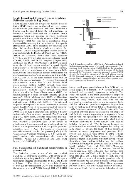
Death Ligand and Receptor Pig Ovarian Follicle 269Death Ligand and Receptor System RegulatesFollicular Atresia <strong>in</strong> Pig OvaryDeath ligands, which are grouped the tumour necrosisfactor (TNF) family, are synthesized as type-II membraneprote<strong>in</strong>s (Ashkenazi and Dixit 1998). Most deathligands can be cleaved from the cell membrane tobecome a soluble form and act as trimers. Deathreceptors, which are classified as type-I membraneprote<strong>in</strong>s, constitute a subfamily with<strong>in</strong> the TNF receptorsuperfamily (TNFRsf) that has a cytoplasmic deathdoma<strong>in</strong> (DD) necessary for the activation of apoptosis(Hengartner 2000). These receptors are trimerized andthen b<strong>in</strong>d to death ligands, which are a trigger forapoptosis. Cell death ligand–receptor systems known <strong>in</strong>mammals <strong>in</strong>clude the Fas ligand (FasL) and Fas (CD95,APO-1 or TNFRsf6), TNFa and TNFa receptors(TNFRs), and TNFa-related apoptosis-<strong>in</strong>duc<strong>in</strong>g ligand(TRAIL, Apo2L) and TRAIL receptors (Nagata 1997;Ashkenazi and Dixit 1998; Wallach et al. 1999). In mostcases, the cell death receptor-mediated apoptotic signall<strong>in</strong>gpathway is as follows: (1) Cell death ligands,b<strong>in</strong>d<strong>in</strong>g with cell membranes or exist<strong>in</strong>g as a solubleform, b<strong>in</strong>d to the extracellular doma<strong>in</strong> of trimerized celldeath receptors, each of which conta<strong>in</strong>s an <strong>in</strong>tracellularDD. (2) The DD of the death receptor b<strong>in</strong>ds with theDD of the adaptor prote<strong>in</strong>s (TNF receptor 1-associateddeath doma<strong>in</strong> prote<strong>in</strong>: TRADD and Fas-associateddeath doma<strong>in</strong> prote<strong>in</strong>: FADD) through homophilic<strong>in</strong>teraction (Inoue et al. 2007). (3) An <strong>in</strong>itiator caspase(procaspase-8) b<strong>in</strong>ds to FADD through homophilic<strong>in</strong>teraction with the death effector doma<strong>in</strong> (DED; theresult<strong>in</strong>g complex is called the ‘death-<strong>in</strong>duc<strong>in</strong>g signall<strong>in</strong>gcomplex’: DISC) (Medema et al. 1997). (4) Dimerizationof procaspase-8 <strong>in</strong>duces auto-proteolytic cleavageand activation (Bold<strong>in</strong> et al. 1995). (5) The activatedcaspase-8 subsequently activates downstream caspaseseither directly (‘type I’) or via mitochondrial perturbation(‘type II; mitochondrion-dependent’) (Matsui et al.2003). (6-a) In type I apoptotic cells, caspase-8 directlyactivates the effector enzyme, caspase-3. (7-a) Trancatedcaspase-3, active form, activates endogenous endonucleasesthat results <strong>in</strong> apoptosis. (6-b) In type II apoptoticcells, caspase-8’s activation leads to the release ofcytochrome c from mitochondrion, which results <strong>in</strong> the<strong>in</strong>teraction of procaspase-9 with apoptotic proteaseactivat<strong>in</strong>gfactor 1 (Apaf1) [formation of cytochromec-Apaf1-caspase-9 complex (apoptosome)]. (7-b) Activatedcaspase-9 cleaves procaspase-3. (8-b) Active caspase-3activates the endonucleases that results <strong>in</strong>apoptosis (Nagata 1997; Ashkenazi and Dixit 1998;Matsui et al. 2003) (Fig. 1).FasL–Fas and other cell death ligand–receptor systems <strong>in</strong>granulosa cellThe FasL–Fas system is one of the most studiedparadigms of <strong>in</strong>structive apoptosis, with strong apoptosis-<strong>in</strong>duc<strong>in</strong>gactivity (Wallach et al. 1999). The apoptosissignal triggered and mediated by FasL and Fas isas follows: (1) FasL b<strong>in</strong>ds to the extracellular doma<strong>in</strong>of Fas. (2) The <strong>in</strong>tracellular DD of Fas <strong>in</strong>teracts withthe adaptor prote<strong>in</strong> (FADD) through DD. (3) FADDDiscFasLFasFADDActivated caspase-8ApoptosisProcaspase-8: Death doma<strong>in</strong> (DD): Death effector doma<strong>in</strong> (DED): Enzymatic doma<strong>in</strong>Cell membraneFig. 1. Intracellular signall<strong>in</strong>g <strong>in</strong> TNF family. When cell death ligandb<strong>in</strong>ds to the extracellular region of cell death receptor, adoptor (Fasassociateddeath doma<strong>in</strong> prote<strong>in</strong>: FADD) b<strong>in</strong>ds to the <strong>in</strong>tracellularregion of receptor through the homophilic <strong>in</strong>teraction of the deathdoma<strong>in</strong>s (DD). Then, <strong>in</strong>itiator caspase (procaspase-8) b<strong>in</strong>ds to FADDthrough the homophilic <strong>in</strong>teraction of the death effector doma<strong>in</strong>(DED). Dimerized procaspase-8 is auto-cleaved, and then truncatedcaspase-8 (active caspase-8) <strong>in</strong>duces activation of down-stream caspases(ex. caspase-3), which results <strong>in</strong> apoptosis<strong>in</strong>teracts with procaspase-8 through their DED and theactive caspase-8 is formed. (4) A caspase cascade isactivated and eventually apoptosis is <strong>in</strong>duced. TheFasL–Fas system is the most characterized apoptosissignall<strong>in</strong>g mach<strong>in</strong>ery <strong>in</strong> granulosa cells (Inoue et al.2006). In many species, both FasL and Fas areexpressed <strong>in</strong> granulosa cells. In mur<strong>in</strong>e ovaries, FasLand Fas mRNA and prote<strong>in</strong> are expressed <strong>in</strong> granulosacells of healthy and atretic follicles (Sakamaki et al.1997). Moreover, treatment of female mice with Fasactivat<strong>in</strong>gantibody promoted granulosa cell apoptosisand follicular atresia, suggest<strong>in</strong>g a pro-apoptotic functionof FasL–Fas signall<strong>in</strong>g <strong>in</strong> vivo. In rat ovaries, FasLand Fas prote<strong>in</strong> occur <strong>in</strong> granulosa cells, which tend tobe more abundant <strong>in</strong> atretic follicles than healthyfollicles (Hakuno et al. 1996). In human females, thegranulosa cells of antral follicles express Fas dur<strong>in</strong>g theearly stages of atresia, the levels of Fas expression<strong>in</strong>crease as atresia progresses, and granulosa FasLprote<strong>in</strong> also <strong>in</strong>creases with atresia <strong>in</strong> antral follicles(Cataldo et al. 2000). In bov<strong>in</strong>e ovaries, FasL mRNAlevels are higher <strong>in</strong> granulosa cells from atretic folliclesthan those from healthy follicles, and Fas mRNAexpression is stronger <strong>in</strong> granulosa cells of atretic thanhealthy follicles (Porter et al. 2000, 2001). Moreover,CD45, a pan-leucocyte marker, was not detected <strong>in</strong>bov<strong>in</strong>e granulosa cells, <strong>in</strong>dicat<strong>in</strong>g that immune cellswere not a source of FasL or Fas <strong>in</strong> the granulosa celllayer (Hu et al. 2001). Trace levels of FasL and FasmRNA and prote<strong>in</strong> were detected <strong>in</strong> granulosa cells ofhealthy follicles but the levels <strong>in</strong>creased dur<strong>in</strong>g atresia<strong>in</strong> porc<strong>in</strong>e ovaries (Inoue et al. 2006). These expressionpatterns of FasL and Fas strongly <strong>in</strong>dicate the importantrole of FasL–Fas signall<strong>in</strong>g <strong>in</strong> follicular atresia.Ó 2008 The Authors. Journal compilation Ó 2008 Blackwell Verlag


