Reproduction in Domestic Animals
Reproduction in Domestic Animals
Reproduction in Domestic Animals
- No tags were found...
You also want an ePaper? Increase the reach of your titles
YUMPU automatically turns print PDFs into web optimized ePapers that Google loves.
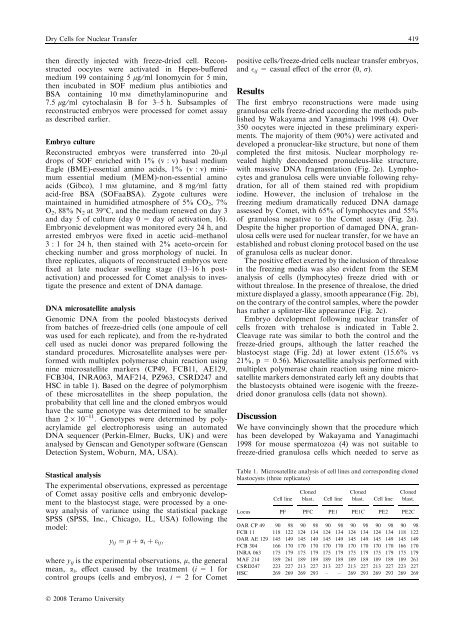
Dry Cells for Nuclear Transfer 419then directly <strong>in</strong>jected with freeze-dried cell. Reconstructedoocytes were activated <strong>in</strong> Hepes-bufferedmedium 199 conta<strong>in</strong><strong>in</strong>g 5 lg ⁄ ml Ionomyc<strong>in</strong> for 5 m<strong>in</strong>,then <strong>in</strong>cubated <strong>in</strong> SOF medium plus antibiotics andBSA conta<strong>in</strong><strong>in</strong>g 10 mM dimethylam<strong>in</strong>opur<strong>in</strong>e and7.5 lg ⁄ ml cytochalas<strong>in</strong> B for 3–5 h. Subsamples ofreconstructed embryos were processed for comet assayas described earlier.Embryo cultureReconstructed embryos were transferred <strong>in</strong>to 20-lldrops of SOF enriched with 1% (v : v) basal mediumEagle (BME)-essential am<strong>in</strong>o acids, 1% (v : v) m<strong>in</strong>imumessential medium (MEM)-non-essential am<strong>in</strong>oacids (Gibco), 1 mM glutam<strong>in</strong>e, and 8 mg ⁄ ml fattyacid-free BSA (SOFaaBSA). Zygote cultures werema<strong>in</strong>ta<strong>in</strong>ed <strong>in</strong> humidified atmosphere of 5% CO 2 ,7%O 2 , 88% N 2 at 39°C, and the medium renewed on day 3and day 5 of culture (day 0 = day of activation, 16).Embryonic development was monitored every 24 h, andarrested embryos were fixed <strong>in</strong> acetic acid–methanol3 : 1 for 24 h, then sta<strong>in</strong>ed with 2% aceto-orce<strong>in</strong> forcheck<strong>in</strong>g number and gross morphology of nuclei. Inthree replicates, aliquots of reconstructed embryos werefixed at late nuclear swell<strong>in</strong>g stage (13–16 h postactivation)and processed for Comet analysis to <strong>in</strong>vestigatethe presence and extent of DNA damage.DNA microsatellite analysisGenomic DNA from the pooled blastocysts derivedfrom batches of freeze-dried cells (one ampoule of cellwas used for each replicate), and from the re-hydratedcell used as nuclei donor was prepared follow<strong>in</strong>g thestandard procedures. Microsatellite analyses were performedwith multiplex polymerase cha<strong>in</strong> reaction us<strong>in</strong>gn<strong>in</strong>e microsatellite markers (CP49, FCB11, AE129,FCB304, INRA063, MAF214, PZ963, CSRD247 andHSC <strong>in</strong> table 1). Based on the degree of polymorphismof these microsatellites <strong>in</strong> the sheep population, theprobability that cell l<strong>in</strong>e and the cloned embryos wouldhave the same genotype was determ<strong>in</strong>ed to be smallerthan 2 · 10 )11 . Genotypes were determ<strong>in</strong>ed by polyacrylamidegel electrophoresis us<strong>in</strong>g an automatedDNA sequencer (Perk<strong>in</strong>-Elmer, Bucks, UK) and wereanalysed by Genscan and Genotyper software (GenscanDetection System, Woburn, MA, USA).Stastical analysisThe experimental observations, expressed as percentageof Comet assay positive cells and embryonic developmentto the blastocyst stage, were processed by a onewayanalysis of variance us<strong>in</strong>g the statistical packageSPSS (SPSS, Inc., Chicago, IL, USA) follow<strong>in</strong>g themodel:y ij ¼ l þ a i þ e ij ;where y ij is the experimental observations, l, the generalmean, a i , effect caused by the treatment (i = 1 forcontrol groups (cells and embryos), i = 2 for Cometpositive cells ⁄ freeze-dried cells nuclear transfer embryos,and ij = casual effect of the error (0, r).ResultsThe first embryo reconstructions were made us<strong>in</strong>ggranulosa cells freeze-dried accord<strong>in</strong>g the methods publishedby Wakayama and Yanagimachi 1998 (4). Over350 oocytes were <strong>in</strong>jected <strong>in</strong> these prelim<strong>in</strong>ary experiments.The majority of them (90%) were activated anddeveloped a pronuclear-like structure, but none of themcompleted the first mitosis. Nuclear morphology revealedhighly decondensed pronucleus-like structure,with massive DNA fragmentation (Fig. 2e). Lymphocytesand granulosa cells were unviable follow<strong>in</strong>g rehydration,for all of them sta<strong>in</strong>ed red with propidiumiod<strong>in</strong>e. However, the <strong>in</strong>clusion of trehalose <strong>in</strong> thefreez<strong>in</strong>g medium dramatically reduced DNA damageassessed by Comet, with 65% of lymphocytes and 55%of granulosa negative to the Comet assay (Fig. 2a).Despite the higher proportion of damaged DNA, granulosacells were used for nuclear transfer, for we have anestablished and robust clon<strong>in</strong>g protocol based on the useof granulosa cells as nuclear donor.The positive effect exerted by the <strong>in</strong>clusion of threalose<strong>in</strong> the freez<strong>in</strong>g media was also evident from the SEManalysis of cells (lymphocytes) freeze dried with orwithout threalose. In the presence of threalose, the driedmixture displayed a glassy, smooth appearance (Fig. 2b),on the contrary of the control samples, where the powderhas rather a spl<strong>in</strong>ter-like appearance (Fig. 2c).Embryo development follow<strong>in</strong>g nuclear transfer ofcells frozen with trehalose is <strong>in</strong>dicated <strong>in</strong> Table 2.Cleavage rate was similar to both the control and thefreeze-dried groups, although the latter reached theblastocyst stage (Fig. 2d) at lower extent (15.6% vs21%, p = 0.56). Microsatellite analysis performed withmultiplex polymerase cha<strong>in</strong> reaction us<strong>in</strong>g n<strong>in</strong>e microsatellitemarkers demonstrated early left any doubts thatthe blastocysts obta<strong>in</strong>ed were isogenic with the freezedrieddonor granulosa cells (data not shown).DiscussionWe have conv<strong>in</strong>c<strong>in</strong>gly shown that the procedure whichhas been developed by Wakayama and Yanagimachi1998 for mouse spermatozoa (4) was not suitable tofreeze-dried granulosa cells which needed to serve asTable 1. Microsatellite analysis of cell l<strong>in</strong>es and correspond<strong>in</strong>g clonedblastocysts (three replicates)LocusCell l<strong>in</strong>eClonedblast.Cell l<strong>in</strong>eClonedblast.Cell l<strong>in</strong>eClonedblast.PF PFC PE1 PE1C PE2 PE2COAR CP 49 90 98 90 98 90 98 90 98 90 98 90 98FCB 11 118 122 124 134 124 134 124 134 124 134 118 122OAR AE 129 145 149 145 149 145 149 145 149 145 149 145 149FCB 304 166 170 170 170 170 170 170 170 170 170 166 170INRA 063 175 179 175 179 175 179 175 179 175 179 175 179MAF 214 189 261 189 189 189 189 189 189 189 189 189 261CSRD247 223 227 213 227 213 227 213 227 213 227 223 227HSC 269 269 269 293 – – 269 293 269 293 269 269Ó 2008 Teramo University


