

Download Complete Issue - Academic Journals
Download Complete Issue - Academic Journals
Download Complete Issue - Academic Journals

Create successful ePaper yourself
Turn your PDF publications into a flip-book with our unique Google optimized e-Paper software.
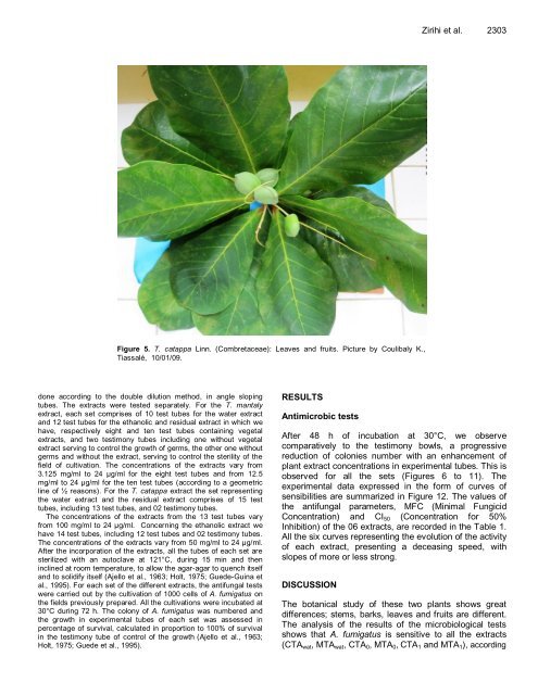
Figure 5. T. catappa Linn. (Combretaceae): Leaves and fruits. Picture by Coulibaly K.,<br />
Tiassalé, 10/01/09.<br />
done according to the double dilution method, in angle sloping<br />
tubes. The extracts were tested separately. For the T. mantaly<br />
extract, each set comprises of 10 test tubes for the water extract<br />
and 12 test tubes for the ethanolic and residual extract in which we<br />
have, respectively eight and ten test tubes containing vegetal<br />
extracts, and two testimony tubes including one without vegetal<br />
extract serving to control the growth of germs, the other one without<br />
germs and without the extract, serving to control the sterility of the<br />
field of cultivation. The concentrations of the extracts vary from<br />
3.125 mg/ml to 24 µg/ml for the eight test tubes and from 12.5<br />
mg/ml to 24 µg/ml for the ten test tubes (according to a geometric<br />
line of ½ reasons). For the T. catappa extract the set representing<br />
the water extract and the residual extract comprises of 15 test<br />
tubes, including 13 test tubes, and 02 testimony tubes.<br />
The concentrations of the extracts from the 13 test tubes vary<br />
from 100 mg/ml to 24 µg/ml. Concerning the ethanolic extract we<br />
have 14 test tubes, including 12 test tubes and 02 testimony tubes.<br />
The concentrations of the extracts vary from 50 mg/ml to 24 µg/ml.<br />
After the incorporation of the extracts, all the tubes of each set are<br />
sterilized with an autoclave at 121°C, during 15 min and then<br />
inclined at room temperature, to allow the agar-agar to quench itself<br />
and to solidify itself (Ajello et al., 1963; Holt, 1975; Guede-Guina et<br />
al., 1995). For each set of the different extracts, the antifungal tests<br />
were carried out by the cultivation of 1000 cells of A. fumigatus on<br />
the fields previously prepared. All the cultivations were incubated at<br />
30°C during 72 h. The colony of A. fumigatus was numbered and<br />
the growth in experimental tubes of each set was assessed in<br />
percentage of survival, calculated in proportion to 100% of survival<br />
in the testimony tube of control of the growth (Ajello et al., 1963;<br />
Holt, 1975; Guede et al., 1995).<br />
RESULTS<br />
Antimicrobic tests<br />
Zirihi et al. 2303<br />
After 48 h of incubation at 30°C, we observe<br />
comparatively to the testimony bowls, a progressive<br />
reduction of colonies number with an enhancement of<br />
plant extract concentrations in experimental tubes. This is<br />
observed for all the sets (Figures 6 to 11). The<br />
experimental data expressed in the form of curves of<br />
sensibilities are summarized in Figure 12. The values of<br />
the antifungal parameters, MFC (Minimal Fungicid<br />
Concentration) and CI50 (Concentration for 50%<br />
Inhibition) of the 06 extracts, are recorded in the Table 1.<br />
All the six curves representing the evolution of the activity<br />
of each extract, presenting a deceasing speed, with<br />
slopes of more or less strong.<br />
DISCUSSION<br />
The botanical study of these two plants shows great<br />
differences; stems, barks, leaves and fruits are different.<br />
The analysis of the results of the microbiological tests<br />
shows that A. fumigatus is sensitive to all the extracts<br />
(CTAwat, MTAwat, CTA0, MTA0, CTA1 and MTA1), according


