Living Image 3.1
Living Image 3.1
Living Image 3.1

Create successful ePaper yourself
Turn your PDF publications into a flip-book with our unique Google optimized e-Paper software.
<strong>Living</strong> <strong>Image</strong> ® Software User’s Manual<br />
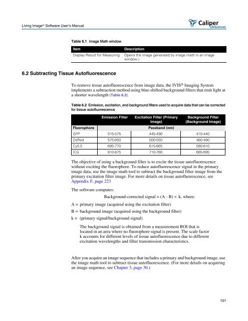
Table 6.1 <strong>Image</strong> Math window<br />
Item Description<br />
6.2 Subtracting Tissue Autofluorescence<br />
Display Result for Measuring Opens the image generated by image math in an image<br />
window.)<br />
To remove tissue autofluorescence from image data, the IVIS ® Imaging System<br />
implements a subtraction method using blue-shifted background filters that emit light at<br />
a shorter wavelength (Table 6.2).<br />
Table 6.2 Emission, excitation, and background filters used to acquire data that can be corrected<br />
for tissue autofluorescence<br />
Emission Filter Excitation Filter (Primary<br />
<strong>Image</strong>)<br />
Fluorophore Passband (nm)<br />
The objective of using a background filter is to excite the tissue autofluorescence<br />
without exciting the fluorophore. To reduce autofluorescence signal in the primary<br />
image data, use the image math tool to subtract the background filter image from the<br />
primary excitation filter image. For more details on tissue autofluorescence, see<br />
Appendix F, page 223<br />
The software computes:<br />
Background-corrected signal = (A - B) × k, where:<br />
A = primary image (acquired using the excitation filter)<br />
B = background image (acquired using the background filter)<br />
k = (primary signal/background signal)<br />
Background Filter<br />
(Background <strong>Image</strong>)<br />
GFP 515-575 445-490 410-440<br />
DsRed 575-650 500-550 460-490<br />
Cy5.5 695-770 615-665 580-610<br />
ICG 810-875 710-760 665-695<br />
The background signal is obtained from a measurement ROI that is<br />
located in an area where no fluorophore signal is present. The scale factor<br />
k accounts for different levels of tissue autofluorescence due to different<br />
excitation wavelengths and filter transmission characteristics.<br />
After you acquire an image sequence that includes a primary and background image, use<br />
the image math tool to subtract tissue autofluorescence. (For more details on acquiring<br />
an image sequence, see Chapter 3, page 30.)<br />
101