Living Image 3.1
Living Image 3.1
Living Image 3.1

You also want an ePaper? Increase the reach of your titles
YUMPU automatically turns print PDFs into web optimized ePapers that Google loves.
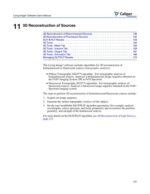
<strong>Living</strong> <strong>Image</strong> ® Software User’s Manual<br />
11<br />
3D Reconstruction of Sources<br />
3D Reconstruction of Bioluminescent Sources . . . . . . . . . . . . . . . . . . . . . . . 138<br />
3D Reconstruction of Fluorescent Sources . . . . . . . . . . . . . . . . . . . . . . . . . 142<br />
DLIT & FLIT Results . . . . . . . . . . . . . . . . . . . . . . . . . . . . . . . . . . . . . . . 144<br />
3D Tools . . . . . . . . . . . . . . . . . . . . . . . . . . . . . . . . . . . . . . . . . . . . . 150<br />
3D Tools - Mesh Tab . . . . . . . . . . . . . . . . . . . . . . . . . . . . . . . . . . . . . . 156<br />
3D Tools - Volume Tab . . . . . . . . . . . . . . . . . . . . . . . . . . . . . . . . . . . . . 158<br />
3D Tools - Organs Tab . . . . . . . . . . . . . . . . . . . . . . . . . . . . . . . . . . . . . 161<br />
3D Tools - Animation Tab . . . . . . . . . . . . . . . . . . . . . . . . . . . . . . . . . . . 168<br />
Managing DLIT/FLIT Results . . . . . . . . . . . . . . . . . . . . . . . . . . . . . . . . . 173<br />
The <strong>Living</strong> <strong>Image</strong> ® software includes algorithms for 3D reconstruction of<br />
bioluminescent or fluorescent sources (tomographic analysis):<br />
• Diffuse Tomography (DLIT) algorithm - For tomographic analysis of<br />
bioluminescent sources. Analyzes a bioluminescent image sequence obtained on<br />
the IVIS ® Imaging System 200 or IVIS Spectrum.<br />
• Fluorescent Tomography (FLIT) algorithm - For tomographic analysis of<br />
fluorescent sources. Analyzes a fluorescent image sequence obtained on the IVIS ®<br />
Spectrum imaging system.<br />
The steps to perform 3D reconstruction of bioluminescent/fluorescent sources include:<br />
1. Acquire an image sequence.<br />
2. Generate the surface topography (surface) of the subject.<br />
3. Set the user-modifiable DLIT/FLIT algorithm parameters (for example, analysis<br />
wavelengths, source spectrum, and tissue properties) and reconstruct the position,<br />
geometry, and strength of the luminescent sources.<br />
For more details on the DLIT/FLIT algorithm, see 3D Reconstruction of Light Sources,<br />
page 233.<br />
137