Living Image 3.1
Living Image 3.1
Living Image 3.1

You also want an ePaper? Increase the reach of your titles
YUMPU automatically turns print PDFs into web optimized ePapers that Google loves.
4. Working With <strong>Image</strong>s<br />
46<br />
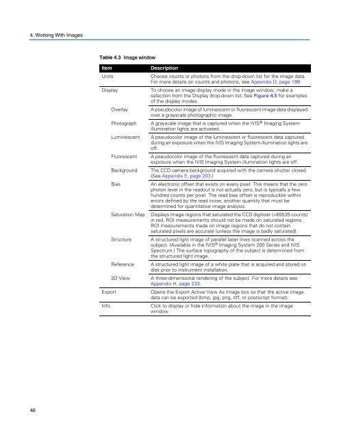
Table 4.3 <strong>Image</strong> window<br />
Item Description<br />
Units Choose counts or photons from the drop-down list for the image data.<br />
For more details on counts and photons, see Appendix D, page 199.<br />
Display To choose an image display mode in the image window, make a<br />
selection from the Display drop-down list. See Figure 4.5 for examples<br />
of the display modes.<br />
Overlay A pseudocolor image of luminescent or fluorescent image data displayed<br />
over a grayscale photographic image.<br />
Photograph A grayscale image that is captured when the IVIS ® Imaging System<br />
illumination lights are activated.<br />
Luminescent A pseudocolor image of the luminescent or fluorescent data captured<br />
during an exposure when the IVIS Imaging System illumination lights are<br />
off.<br />
Fluorescent A pseudocolor image of the fluorescent data captured during an<br />
exposure when the IVIS Imaging System illumination lights are off.<br />
Background The CCD camera background acquired with the camera shutter closed.<br />
(See Appendix E, page 203.)<br />
Bias An electronic offset that exists on every pixel. This means that the zero<br />
photon level in the readout is not actually zero, but is typically a few<br />
hundred counts per pixel. The read bias offset is reproducible within<br />
errors defined by the read noise, another quantity that must be<br />
determined for quantitative image analysis.<br />
Saturation Map Displays image regions that saturated the CCD digitizer (>65535 counts)<br />
in red. ROI measurements should not be made on saturated regions.<br />
ROI measurements made on image regions that do not contain<br />
saturated pixels are accurate (unless the image is badly saturated).<br />
Structure A structured light image of parallel laser lines scanned across the<br />
subject. (Available in the IVIS ® Imaging System 200 Series and IVIS<br />
Spectrum.) The surface topography of the subject is determined from<br />
the structured light image.<br />
Reference A structured light image of a white plate that is acquired and stored on<br />
disk prior to instrument installation.<br />
3D View A three-dimensional rendering of the subject. For more details see<br />
Appendix H, page 233.<br />
Export Opens the Export Active View As <strong>Image</strong> box so that the active image<br />
data can be exported (bmp, jpg, png, tiff, or postscript format).<br />
Info Click to display or hide information about the image in the image<br />
window.