Online proceedings - EDA Publishing Association
Online proceedings - EDA Publishing Association
Online proceedings - EDA Publishing Association

You also want an ePaper? Increase the reach of your titles
YUMPU automatically turns print PDFs into web optimized ePapers that Google loves.
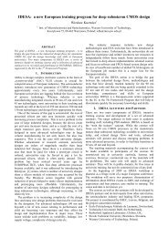
Fig. 5 Computed absolute temperature contours, T (°C), at the<br />
surface level of the polysilicon resistor for P = 250 mW<br />
activation power. Maximum computed temperature is 69.8°C<br />
(the reference temperature was 20 °C and the shown area is a<br />
54 by 54 μm square portion of the domain)<br />
C. Validation of temperature results: numerical simulation<br />
results<br />
The experimental results obtained here were validated<br />
using the authors’ ultra-fast self-adaptive numerical<br />
simulation engine [8,9] A model was built based on the<br />
device design data from Austria Microsystems and computed<br />
using our solver with the results presented in Fig. 5, for an<br />
activation power of 250 mW. The power was considered to<br />
be uniform and covers the entire area of the polysilicon<br />
resistor. The temperature presented in Fig. 5 is the absolute<br />
temperature in °C; thus, to obtain the temperature change,<br />
the reference temperature of 20°C should be subtracted from<br />
this value. The computed maximum temperature difference<br />
of 69.8°C agrees well, within 5%, with the experimentally<br />
measured temperature plotted in Fig. 4.<br />
D. Validation of temperature results: built-in diode results<br />
In addition to comparing the experimental results to the<br />
results of the numerical simulation, the results are also<br />
Fig. 6 Temperature measured using the built-in diode versus<br />
applied electrical power to the polysilicon resistor.<br />
7-9 October 2009, Leuven, Belgium<br />
checked against temperature readings obtained from an<br />
embedded temperature sensor. A diode was used to measure<br />
the temperature which is located in the center region of the<br />
C-shaped microresistor.<br />
First, the diode was calibrated using our thermoelectric<br />
element based stage by measuring the change in the voltage<br />
value with the change in the base temperature while the<br />
current was kept constant at 1 μA. It was found that the<br />
voltage decreases linearly with the temperature at a rate of<br />
2.55 mV/°C for the range of temperature considered here.<br />
After calibrating the diode, the polysilicon resistor was<br />
activated at various power levels and the voltage of the diode<br />
was recorded while again keeping the diode current constant<br />
at 1 μA. The obtained data is plotted in Fig. 6 and shows<br />
that the diode voltage is linearly proportional to the applied<br />
microresistor power. For the 250 mW of applied power, the<br />
diode reads a temperature gradient of 23.5°C which grossly<br />
underestimates the computed 69.8°C maximum temperature<br />
obtained for the polysilicon resistor. Nevertheless, by<br />
investigating both the experimental results shown in Fig. 4<br />
and the numerical results presented in Fig. 5 one might find<br />
out that indeed the temperature at the center location of the<br />
poly resistor is expected to be much lower than the<br />
maximum temperature observed on the surface of the<br />
resistor itself. Therefore, we must conclude that the<br />
embedded diode sensor may not be as useful as initially<br />
thought at determining the average temperature of the poly<br />
resistor.<br />
III. CONCLUSIONS<br />
This work presented for the first time a successful method<br />
for calibrating pixel-by-pixel and in-situ a CCD camerabased<br />
thermoreflectance thermography system with<br />
nanometer spatial resolution. This article described the<br />
measurement system and methodology used and presents<br />
relevant results. Using the thermoreflectance method to<br />
determine the temperature map of an activated device<br />
requires two steps: first, the thermal image is acquired using<br />
a CCD camera and, second, the obtained thermal image is<br />
converted to the actual temperature map by multiplying each<br />
pixel of the thermal image with the corresponding<br />
thermoreflectance coefficient. The critical aspect is that even<br />
if the thermoreflectance coefficient is known, or measured<br />
independently for each material present on the surface of the<br />
sample, converting the thermal image to the temperature<br />
map requires building manually the exact corresponding map<br />
of the thermoreflectance coefficient, which in the case of<br />
complex microelectronic devices might be difficult. In<br />
addition, it turns out that the thermoreflectance coefficient is<br />
highly dependent on the wavelength of the probing light,<br />
numerical aperture, focus level, light uniformity, and other<br />
measurement effects. To mitigate these issues, one must<br />
obtain the thermoreflectance coefficient map of the same<br />
measurement area of interest, while ensuring that the same<br />
objective lens is used, the same focus level is maintained,<br />
and the same exact position is kept for frame acquisition at<br />
both the low and high temperature settings. To satisfy all of<br />
these requirements, the position of the sample must be<br />
adjusted in 3D space with nanometer spatial resolution.<br />
©<strong>EDA</strong> <strong>Publishing</strong>/THERMINIC 2009 134<br />
ISBN: 978-2-35500-010-2