Table of Contents - WOC 2012
Table of Contents - WOC 2012
Table of Contents - WOC 2012

Create successful ePaper yourself
Turn your PDF publications into a flip-book with our unique Google optimized e-Paper software.
Computer-Aided Diagnosis, Novel Image Processing and Analysis<br />
Sat 18 Feb 13:30 - 15:00 Conference Room B2<br />
IS-TEL-SA 213 (1)<br />
Low Cost Fundus Camera-System for Telemedical Applications<br />
Hoeher Bernhard (1, 3, 5) , Schmauss Bernhard (1, 3, 4, 5) (2, 3, 4)<br />
, Michelson Georg<br />
1. Microwave Engineering, University <strong>of</strong> Erlangen-Nuremberg<br />
2. Department <strong>of</strong> Ophthalmology, University <strong>of</strong> Erlangen-Nuremberg<br />
3. Graduate School in Advanced Optical Technologies (SAOT)<br />
4. Interdisciplinary Center for Ophthalmological Preventive Medicine and Imaging<br />
5. Medical Valley EMN, Leading-Edge Cluster<br />
Medical examinations in ophthalmology <strong>of</strong>ten require expensive devices.<br />
Therefore many people living in regions with insufficient health care facilities<br />
cannot be treated for eye diseases due to missing medical equipment and for<br />
lack <strong>of</strong> qualified eye doctors. Hence we design a low cost, robust and highly<br />
automated non-mydriatic fundus camera-system with a telemedical interface.<br />
Thereby fundus images can be taken even by less qualified people and<br />
the diagnosis is done by specialists located elsewhere. The fundus images<br />
are transmitted by the local mobile phone network. We take advantage<br />
<strong>of</strong> low prices in three high-tech sectors: Optics for amateur astronomy and<br />
photography, computer hardware and mobile phone technology. We embed<br />
technology from all three sectors in one powerful system. We found that even<br />
cheap eyepieces together with a one mega pixel camera give fundus images<br />
<strong>of</strong> sufficient quality. In addition we increased performance by applying image<br />
processing technologies. The fundus camera will be capable to screen a large<br />
number <strong>of</strong> people. For the eye doctors this means an immense time saving by<br />
just clicking through fundus images quickly. Thus many people can effectively<br />
be examined, even though there is no expert on site.<br />
IS-TEL-SA 213 (2)<br />
The Erlangen Glaucoma Matrix<br />
Hornegger Joachim (1,2) , Meier Jörg (1,2) , Bock Rüdiger (1,2) ,<br />
Michelson Georg (2,3,4)<br />
1. School <strong>of</strong> Advanced Optical Technologies, University Erlangen-Nuremberg<br />
2. Pattern Recognition Lab, University Erlangen-Nuremberg<br />
3. Online Journals <strong>of</strong> Ophthalmology<br />
4. Interdisciplinary Center <strong>of</strong> Ophthalmic Preventive Medicine and Imaging, University<br />
Erlangen-Nuremberg<br />
Background and Purpose: Glaucoma is one <strong>of</strong> the most common causes for<br />
blindness worldwide. The early and reliable diagnosis <strong>of</strong> glaucoma is important<br />
to slow down the progression <strong>of</strong> this particular disease. Efficient, effective<br />
and low-cost screening methods are required but still do not exist. Research<br />
focuses both on concepts for low-cost fundus cameras, and algorithmic tools<br />
for computer aided diagnosis <strong>of</strong> fundus images. In this contribution a twodimensional<br />
visualization tool is introduced that allows for the comparison <strong>of</strong><br />
the current image with pre-diagnosed image data. The automated computation<br />
<strong>of</strong> a glaucoma risk index on color fundus photographs is used to spatially<br />
assign an undiagnosed image to a point in the two-dimensional space <strong>of</strong><br />
reference data.<br />
Methods: The chosen two-stage approach computes in the first step the<br />
probability for glaucoma disease based on a single color fundus image, and<br />
in a second step the acquired image is visualized in a matrix where the matrix<br />
components are pre-diagnosed reference images. The matrix separates, for<br />
instance, different papilla sizes (columns) and varying stages <strong>of</strong> the glaucoma<br />
disease (rows), but any other feature types can be used for the visualization<br />
<strong>of</strong> topological structures <strong>of</strong> image similarities in two dimensions. The reference<br />
image database holds papilla-centered color fundus photographs (Kowa nonmyd,<br />
FOV 22.5°) from the Erlangen Glaucoma Registry (EGR). The gold<br />
standard for glaucoma diagnosis is defined by the diagnosis <strong>of</strong> experienced<br />
ophthalmologists using in addition to the fundus image the following tools:<br />
ophthalmoscopy, visual field test, IOP, FDT and HRT II.<br />
Results: SVM-based glaucoma classification <strong>of</strong> PCA-transformed pixel<br />
intensities leads to an accuracy <strong>of</strong> 0.83 (specificity: 0.72, sensitivity: 0.94,<br />
ROC area: 0.90). Using coefficients <strong>of</strong> spline functions instead <strong>of</strong> PCA features<br />
leads to a recognition rate <strong>of</strong> 0.86 (specificity: 0.78, sensitivity: 0.94, ROC<br />
area: 0.88). The combination <strong>of</strong> both features increases the area under the<br />
ROC curve to 0.93. Also specificity slightly increased to 0.82, and sensitivity<br />
lead to 0.92. The proposed visualization allows evaluating an image in the<br />
context <strong>of</strong> given pre-diagnosed reference samples. Due to the two-dimensional<br />
presentation, non-disease- dependent variations (papilla size, illumination,<br />
etc.) and glaucomatous changes are shown separately.<br />
Conclusions: The experimental evaluation shows that fundus images are a<br />
solid base for computer-assisted glaucoma screening, if modern pattern<br />
recognition technology is applied. The chosen approach gives insights on<br />
glaucomatous optic nerve appearance in relation to varying papilla sizes as<br />
it shows a large diagnosed set <strong>of</strong> data separate for different glaucoma stages<br />
and papilla sizes.<br />
<strong>WOC</strong><strong>2012</strong> Abstract Book<br />
IS-TEL-SA 213 (3)<br />
Advanced Fusion Based Analysis <strong>of</strong> Ophthalmological Image Data<br />
Jan Jiri (1) , Kolar R. (1) , Odstrcilik J. (1) , Gazarek J. (1)<br />
1. Brno University <strong>of</strong> Technology<br />
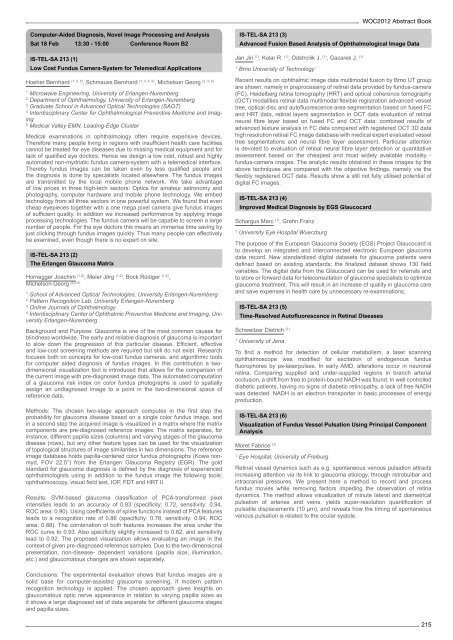
Recent results on ophthalmic image data multimodal fusion by Brno UT group<br />
are shown, namely in preprocessing <strong>of</strong> retinal data provided by fundus-camera<br />
(FC), Heidelberg retina tomography (HRT) and optical coherence tomography<br />
(OCT) modalities retinal data multimodal flexible registration advanced vessel<br />
tree, optical disc and aut<strong>of</strong>luorescence area segmentation based on fused FC<br />
and HRT data, retinal layers segmentation in OCT data evaluation <strong>of</strong> retinal<br />
neural fibre layer based on fused FC and OCT data: combined results <strong>of</strong><br />
advanced texture analysis in FC data compared with registered OCT 3D data<br />
high resolution retinal FC image database with medical expert evaluated vessel<br />
tree segmentations and neural fibre layer assessment. Particular attention<br />
is devoted to evaluation <strong>of</strong> retinal neural fibre layer detection or quantitative<br />
assessment based on the cheapest and most widely available modality -<br />
fundus-camera images. The analytic results obtained in these images by the<br />
above techniques are compared with the objective findings, namely via the<br />
flexibly registered OCT data. Results show a still not fully utilised potential <strong>of</strong><br />
digital FC images.<br />
IS-TEL-SA 213 (4)<br />
Improved Medical Diagnosis by EGS Glaucocard<br />
Schargus Marc (1) , Grehn Franz<br />
1. University Eye Hospital Wuerzburg<br />
The purpose <strong>of</strong> the European Glaucoma Society (EGS) Project Glaucocard is<br />
to develop an integrated and interconnected electronic European glaucoma<br />
data record. New standardized digital datasets for glaucoma patients were<br />
defined based on existing standards; the finalized dataset shows 130 field<br />
variables. The digital data from the Glaucocard can be used for referrals and<br />
to store or forward data for teleconsultation <strong>of</strong> glaucoma specialists to optimize<br />
glaucoma treatment. This will result in an increase <strong>of</strong> quality in glaucoma care<br />
and save expenses in health care by unnecessary re-examinations.<br />
IS-TEL-SA 213 (5)<br />
Time-Resolved Aut<strong>of</strong>luorescence in Retinal Diseases<br />
Schweitzer Dietrich (1)<br />
1. University <strong>of</strong> Jena<br />
To find a method for detection <strong>of</strong> cellular metabolism, a laser scanning<br />
ophthalmoscope was modified for excitation <strong>of</strong> endogenous fundus<br />
fluorophores by ps-laserpulses. In early AMD, alterations occur in neuronal<br />
retina. Comparing supplied and under-supplied regions in branch arterial<br />
occlusion, a shift from free to protein-bound NADH was found. In well controlled<br />
diabetic patients, having no signs <strong>of</strong> diabetic retinopathy, a lack <strong>of</strong> free NADH<br />
was detected. NADH is an electron transporter in basic processes <strong>of</strong> energy<br />
production.<br />
IS-TEL-SA 213 (6)<br />
Visualization <strong>of</strong> Fundus Vessel Pulsation Using Principal Component<br />
Analysis<br />
Moret Fabrice (1)<br />
1. Eye Hospital, University <strong>of</strong> Freiburg<br />
Retinal vessel dynamics such as e.g. spontaneous venous pulsation attracts<br />
increasing attention via its link to glaucoma etiology, through retrobulbar and<br />
intracranial pressures. We present here a method to record and process<br />
fundus movies while removing factors impeding the observation <strong>of</strong> retina<br />
dynamics. The method allows visualization <strong>of</strong> minute lateral and diametrical<br />
pulsation <strong>of</strong> arteries and veins, yields super-resolution quantification <strong>of</strong><br />
pulsatile displacements (10 µm), and reveals how the timing <strong>of</strong> spontaneous<br />
venous pulsation is related to the ocular systole.<br />
215