Annual Meeting Proceedings Part 1 - American Society of Clinical ...
Annual Meeting Proceedings Part 1 - American Society of Clinical ...
Annual Meeting Proceedings Part 1 - American Society of Clinical ...
You also want an ePaper? Increase the reach of your titles
YUMPU automatically turns print PDFs into web optimized ePapers that Google loves.
554s Melanoma/Skin Cancers<br />
8556 General Poster Session (Board #33F), Sun, 8:00 AM-12:00 PM<br />
Everolimus in combination with paclitaxel and carboplatin in patients with<br />
advanced melanoma: A phase II trial <strong>of</strong> the Sarah Cannon Research<br />
Institute (SCRI). Presenting Author: Ralph J. Hauke, Nebraska Cancer<br />
Specialists, Omaha, NE<br />
Background: The PI3k/AKT pathway is activated in most metastatic melanomas;<br />
mTOR is a critical component <strong>of</strong> this pathway. Everolimus, an mTOR<br />
inhibitor, has demonstrated single-agent activity in patients with advanced<br />
melanoma. We evaluated the efficacy and toxicity <strong>of</strong> everolimus in<br />
combination with paclitaxel/carboplatin in patients with advanced melanoma.<br />
Methods: Eligible patients had stage IV or unresectable stage III<br />
melanoma, unselected for braf status, previously untreated with chemotherapy<br />
or targeted agents. Previous immunotherapy was allowed. Additional<br />
eligibility criteria: ECOG PS 0 or 1; measurable disease; no active<br />
brain metastases; adequate bone marrow, kidney, and liver function;<br />
informed consent. All patients received paclitaxel 175mg/m2 , 1-3 hour IV<br />
infusion, and carboplatin AUC 6.0 IV on day 1 <strong>of</strong> each 21-day cycle.<br />
Everolimus 5mg PO was given daily. Patients were evaluated for response<br />
every 6 weeks; treatment continued until progression or undue toxicity.<br />
Median progression-free survival (PFS) for paclitaxel/carboplatin treatment<br />
is 4 months; we looked for a median PFS <strong>of</strong> 6 months with this novel<br />
combination. Results: Seventy patients were treated between 2/2010 and<br />
2/2011; median age 63, 90% had stage IV melanoma. 91% <strong>of</strong> patients<br />
received at least 2 cycles <strong>of</strong> therapy; median cycles received: 4 (range:<br />
1-25�). Twelve patients (17%) had partial responses; an additional 42<br />
patients (60%) had stable disease at first reevaluation. After a median 13<br />
months <strong>of</strong> followup, the median PFS for the entire group was 4 months<br />
(95% CI: 2.8 – 5.0 months); 96% had progressed during the first 12<br />
months. Median survival was 10 months (95% CI: 7.3 – 10.9 months).<br />
Toxicity was as previously described with these agents; neutropenia was the<br />
most common grade 3/4 toxicity (27%). Only 3 patients stopped treatment<br />
due to toxicity. Conclusions: The addition <strong>of</strong> everolimus to paclitaxel/<br />
carboplatin was feasible and well-tolerated; however, efficacy results were<br />
similar to those reported with paclitaxel/carboplatin alone. Further development<br />
<strong>of</strong> this combination regimen for treatment <strong>of</strong> metastatic melanoma is<br />
not recommended.<br />
8558 General Poster Session (Board #33H), Sun, 8:00 AM-12:00 PM<br />
Patterns <strong>of</strong> progression in patients (pts) with V600 BRAF-mutated melanoma<br />
metastatic to the brain treated with dabrafenib (GSK2118436).<br />
Presenting Author: Mary W. F. Azer, The Crown Princess Mary Cancer<br />
Centre Westmead, Sydney, Australia<br />
Background: Dabrafenib has shown efficacy in pts with previously untreated<br />
brain metastases (BM) but most will progress. We report the pattern <strong>of</strong><br />
disease progression (PD) in pts with either previously treated (surgery (Sx),<br />
SRS, WBRT) or untreated BM on dabrafenib. Methods: Clinicopathologic<br />
parameters were collected on 23 pts enrolled in the brain cohort <strong>of</strong> the<br />
BRF112680 phase I and BRF113929 phase II BM study <strong>of</strong> dabrafenib at<br />
Westmead Hospital between Sept 2009 and June 2011. Pts with RECIST<br />
PD but ongoing clinical benefit were allowed to continue dabrafenib.<br />
Results: 12 pts (52%) had previously untreated BM and 11 (48%) were<br />
previously treated with evidence <strong>of</strong> progression prior to dabrafenib. Median<br />
OS from study entry (8.4 mo, 95% CI 5.0-11.8), diagnosis <strong>of</strong> first BM<br />
(11.0 mo, 95% CI 9.1-13.0), and stage IV diagnosis (17.5 mo, 95% CI<br />
12.1-23.0) were not different between the two groups. Similarly, PFS did<br />
not differ between groups. Of the 19 pts who had RECIST PD at datacut, 13<br />
pts had intracranial (IC) PD with no pts progressing in new brain lesions<br />
alone (see table). At IC PD, 3/13 underwent SRS/Sx, 5/13 WBRT and 5/13<br />
had no salvage local therapy to BM. 8 pts continued dabrafenib beyond IC<br />
PD, median 36 days; range 28-273. All measures <strong>of</strong> baseline disease<br />
burden correlated with worse OS; elevated LDH (HR 1.003, 95% CI<br />
1.0-1.007, P�0.044), increased number (no.) metastatic sites (HR 1.32,<br />
95% CI 0.97-1.81, P�0.08), increased no. <strong>of</strong> IC lesions (HR 1.04, 95%CI<br />
1.00-1.09, P�0.04), increased no. extracranial (EC) lesions (HR 1.07,<br />
95%CI 1.02-1.12, P�0.01) and increased RECIST sum <strong>of</strong> diameters<br />
(SoD) (HR 1.012, 95% CI 1.003-1.021, P�0.007). High baseline SoD<br />
was the only factor that predicted worse RECIST response. Conclusions:<br />
<strong>Clinical</strong> benefit from dabrafenib does not appear to be influenced by prior<br />
local therapies to BM. There was no dominant pattern <strong>of</strong> progression in pts<br />
with BM on dabrafenib, but PD due to new lesions alone is rare. Pts with IC<br />
PD may benefit from salvage local therapy and continued dabrafenib.<br />
Extent and burden <strong>of</strong> disease correlates with reduced OS, but not RECIST<br />
response.<br />
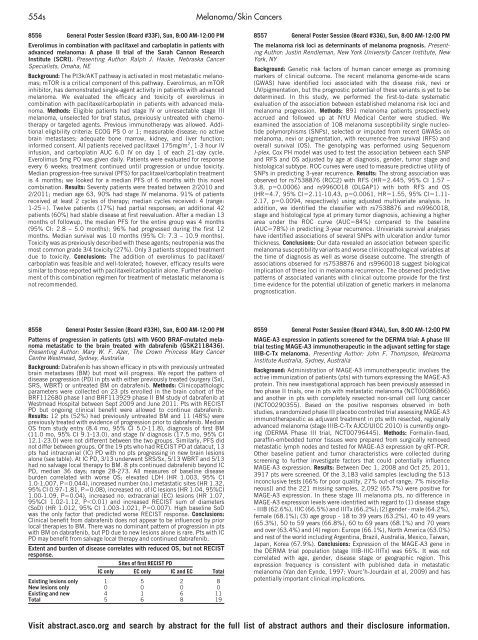
Sites <strong>of</strong> first RECIST PD<br />
IC only EC only IC and EC Total<br />
Existing lesions only 1 5 2 8<br />
New lesions only 0 0 0 0<br />
Existing and new 4 1 6 11<br />
Total 5 6 8 19<br />
8557 General Poster Session (Board #33G), Sun, 8:00 AM-12:00 PM<br />
The melanoma risk loci as determinants <strong>of</strong> melanoma prognosis. Presenting<br />
Author: Justin Rendleman, New York University Cancer Institute, New<br />
York, NY<br />
Background: Genetic risk factors <strong>of</strong> human cancer emerge as promising<br />
markers <strong>of</strong> clinical outcome. The recent melanoma genome-wide scans<br />
(GWAS) have identified loci associated with the disease risk, nevi or<br />
UV/pigmentation, but the prognostic potential <strong>of</strong> these variants is yet to be<br />
determined. In this study, we performed the first-to-date systematic<br />
evaluation <strong>of</strong> the association between established melanoma risk loci and<br />
melanoma progression. Methods: 891 melanoma patients prospectively<br />
accrued and followed up at NYU Medical Center were studied. We<br />
examined the association <strong>of</strong> 108 melanoma susceptibility single nucleotide<br />
polymorphisms (SNPs), selected or imputed from recent GWASs on<br />
melanoma, nevi or pigmentation, with recurrence-free survival (RFS) and<br />
overall survival (OS). The genotyping was performed using Sequenom<br />
I-plex. Cox PH model was used to test the association between each SNP<br />
and RFS and OS adjusted by age at diagnosis, gender, tumor stage and<br />
histological subtype. ROC curves were used to measure predictive utility <strong>of</strong><br />
SNPs in predicting 3-year recurrence. Results: The strong association was<br />
observed for rs7538876 (RCC2) with RFS (HR�2.445, 95% CI 1.57 –<br />
3.8, p�0.0006) and rs9960018 (DLGAP1) with both RFS and OS<br />
(HR�4.7, 95% CI�2.11-10.43, p�0.0061, HR�1.55, 95% CI�1.11-<br />
2.17, p�0.0094, respectively) using adjusted multivariate analysis. In<br />
addition, we identified the classifier with rs7538876 and rs9960018,<br />
stage and histological type at primary tumor diagnosis, achieving a higher<br />
area under the ROC curve (AUC�84%) compared to the baseline<br />
(AUC�78%) in predicting 3-year recurrence. Univariate survival analyses<br />
have identified associations <strong>of</strong> several SNPs with ulceration and/or tumor<br />
thickness. Conclusions: Our data revealed an association between specific<br />
melanoma susceptibility variants and worse clinicopathological variables at<br />
the time <strong>of</strong> diagnosis as well as worse disease outcome. The strength <strong>of</strong><br />
associations observed for rs7538876 and rs9960018 suggest biological<br />
implication <strong>of</strong> these loci in melanoma recurrence. The observed predictive<br />
patterns <strong>of</strong> associated variants with clinical outcome provide for the first<br />
time evidence for the potential utilization <strong>of</strong> genetic markers in melanoma<br />
prognostication.<br />
8559 General Poster Session (Board #34A), Sun, 8:00 AM-12:00 PM<br />
MAGE-A3 expression in patients screened for the DERMA trial: A phase III<br />
trial testing MAGE-A3 immunotherapeutic in the adjuvant setting for stage<br />
IIIB-C-Tx melanoma. Presenting Author: John F. Thompson, Melanoma<br />
Institute Australia, Sydney, Australia<br />
Background: Administration <strong>of</strong> MAGE-A3 immunotherapeutic involves the<br />
active immunization <strong>of</strong> patients (pts) with tumors expressing the MAGE-A3<br />
protein. This new investigational approach has been previously assessed in<br />
two phase II trials, one in pts with metastatic melanoma (NCT00086866)<br />
and another in pts with completely resected non-small cell lung cancer<br />
(NCT00290355). Based on the positive responses observed in both<br />
studies, a randomized phase III placebo controlled trial assessing MAGE-A3<br />
immunotherapeutic as adjuvant treatment in pts with resected, regionally<br />
advanced melanoma (stage IIIB-C-Tx AJCC/UICC 2010) is currently ongoing<br />
(DERMA Phase III trial, NCT00796445). Methods: Formalin-fixed,<br />
paraffin-embedded tumor tissues were prepared from surgically removed<br />
metastatic lymph nodes and tested for MAGE-A3 expression by qRT-PCR.<br />
Other baseline patient and tumor characteristics were collected during<br />
screening to further investigate factors that could potentially influence<br />
MAGE-A3 expression. Results: Between Dec 1, 2008 and Oct 25, 2011,<br />
3917 pts were screened. Of the 3,183 valid samples (excluding the 513<br />
inconclusive tests [66% for poor quality, 27% out-<strong>of</strong> range, 7% miscellaneous])<br />
and the 221 missing samples, 2,092 (65.7%) were positive for<br />
MAGE-A3 expression. In these stage III melanoma pts, no difference in<br />
MAGE-A3 expression levels were identified with regard to (1) disease stage<br />
- IIIB (62.6%), IIIC (66.5%) and IIITx (66.2%); (2) gender - male (64.2%),<br />
female (68.1%); (3) age group - 18 to 39 years (63.2%), 40 to 49 years<br />
(65.3%), 50 to 59 years (66.8%), 60 to 69 years (68.1%) and 70 years<br />
and over (63.4%) and (4) region: Europe (66.1%), North America (63.0%)<br />
and rest <strong>of</strong> the world including Argentina, Brazil, Australia, Mexico, Taiwan,<br />
Japan, Korea (67.9%). Conclusions: Expression <strong>of</strong> the MAGE-A3 gene in<br />
the DERMA trial population (stage IIIB-IIIC-IIITx) was 66%. It was not<br />
correlated with age, gender, disease stage or geographic region. This<br />
expression frequency is consistent with published data in metastatic<br />
melanoma (Van den Eynde, 1997; Vourc’h-Jourdain et al, 2009) and has<br />
potentially important clinical implications.<br />
Visit abstract.asco.org and search by abstract for the full list <strong>of</strong> abstract authors and their disclosure information.


