

Annual Meeting Proceedings Part 1 - American Society of Clinical ...
Annual Meeting Proceedings Part 1 - American Society of Clinical ...
Annual Meeting Proceedings Part 1 - American Society of Clinical ...

Create successful ePaper yourself
Turn your PDF publications into a flip-book with our unique Google optimized e-Paper software.
564s Melanoma/Skin Cancers<br />
8596 General Poster Session (Board #38F), Sun, 8:00 AM-12:00 PM<br />
Detection <strong>of</strong> BRAF mutations in melanoma: Rate <strong>of</strong> mutation detection at<br />
codon 600 using Sanger sequencing as compared to the cobas 4800<br />
method. Presenting Author: Kevin Z. Qu, Nichols Institute, San Juan<br />
Capistrano, CA<br />
Background: Detection <strong>of</strong> BRAF V600 mutations is currently a prerequisite<br />
for approved use <strong>of</strong> vemurafenib in patients with metastatic melanoma. The<br />
cobas 4800 BRAF V600 Mutation Test (Roche Molecular Diagnostics), a<br />
PCR-based assay approved to aid in selecting patients for vemurafenib<br />
therapy, primarily detects V600E. It is also reported to detect V600K,<br />
which has been associated with vemurafenib response as well. We<br />
compared the mutation detection rate <strong>of</strong> the cobas assay with that <strong>of</strong><br />
Sanger sequencing. Methods: 125 de-identified FFPE tissues submitted for<br />
BRAF mutation analysis that all showed histologically-confirmed melanoma<br />
were tested. BRAF mutations were detected using both the cobas kit<br />
and bidirectional Sanger sequencing using BigDye kits (Applied Biosystems).<br />
DNA was extracted from 5-um sections without macrodissection<br />
using the cobas DNA extraction kit (for the cobas test) or from 5-10-um<br />
sections using Agencourt extraction kits (Beckman Coulter) following<br />
macrodissection. Results: The two methods showed agreement in 104/125<br />
(83.2%) <strong>of</strong> cases (Table). Sanger sequencing detected V600 dinucleotide<br />
mutations in 9 samples that were negative by the cobas assay. Sanger<br />
sequencing produced no results in 10 cases owing to suboptimal PCR,<br />
including 2 that were positive by the cobas assay. The cobas assay<br />
produced 2 invalid results, including 1 that was positive for V600E by<br />
Sanger.The cobas assay detected 7/11 V600K mutations. Conclusions:<br />
Overall agreement between cobas and Sanger sequencing was 83.2%. The<br />
Sanger method had higher analytic sensitivity, resulting in nine additional<br />
V600 mutations not called by cobas compared to the two seen by cobas but<br />
not Sanger sequencing. Thus, 16% (9/57) more patients would be<br />
identified as candidates for vemurafenib therapy using the Sanger method.<br />
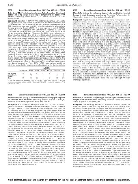
Sanger<br />
sequencing<br />
Not detected V600E V600K V600R Non-600 No result Total<br />
Not detected 56 3 a<br />
4 b<br />
2 c<br />
2 d<br />
7 74<br />
cobas detected 40 7 2 49<br />
Invalid 1 1 2<br />
Total 56 44 11 2 2 10 125<br />
a 2/3 had dinucleotide mutations (GTG�GAA) and 1 had V600_K601del. b All had<br />
dinucleotide mutations (GTG�AAG). c Both had GTG�AGG dinucleotide mutations. d 1)<br />
G469R; 2) E501G.<br />
8598 General Poster Session (Board #38H), Sun, 8:00 AM-12:00 PM<br />
Pharmacodynamic activity <strong>of</strong> selumetinib to predict radiographic response<br />
in advanced uveal melanoma. Presenting Author: Richard D. Carvajal,<br />
Memorial Sloan-Kettering Cancer Center, New York, NY<br />
Background: Functionally activating mutations (mut) in Gnaq or Gna11,<br />
genes that encode for widely expressed G-protein alpha subunits, are early<br />
oncogenic events in uveal melanoma (UM) development and result in<br />
activation <strong>of</strong> the MAPK pathway. We previously demonstrated effective<br />
pathway inhibition with selumetinib (AZD6244, ARRY-142866) in UM cell<br />
lines, with decreased viability associated with pERK and cyclinD1 suppression<br />
(Ambrosini, AACR 2010). Methods: Using paired metastatic tumor<br />
biopsies from patients (pts) with radiographically progressing UM treated<br />
with selumetinib 75 mg BID on a phase II trial (NCT01143402), we<br />
correlated MAPK pathway inhibition with radiographic tumor regression<br />
and clinical benefit. Biopsies were performed at baseline and after 14 �/-1<br />
days <strong>of</strong> treatment. Western blotting was performed for pERK and cyclinD1,<br />
and quantitated by densitometry. Response (RECIST 1.1) was assessed at<br />
baseline, week (wk) 4, wk 8, and q8wks subsequently. Radiographic<br />
regression was defined as greatest percentage shrinkage from baseline.<br />
<strong>Clinical</strong> benefit was defined as RECIST response or stable disease �16wks.<br />
Results: Paired tumor biopsies were assayed from 18 pts: median age 60<br />
(range 47-81), M:F 11:7, median 1 prior therapy (range 0-2), 17 with liver<br />
involvement, Gnaq mut:Gna11 mut:wild-type 8:9:1. Radiographic regression<br />
was observed in 5 pts, with 2 achieving partial responses. 4 pts were<br />
on study �16wks (16�, 20, 25, 31 wks), with one currently on study at<br />
11� wks. Median pERK and cyclinD1 as measured by densitometry<br />
decreased by 48% (p�.03) and 76% (p�.03), respectively. Radiographic<br />
regression correlated with suppression <strong>of</strong> pERK (Spearmen’s rank correlation;<br />
p�0.04) but not cyclinD1 (p�0.38). A trend towards pERK suppression<br />
correlating with clinical benefit was observed (p�.07) with each <strong>of</strong> the<br />
5 pts achieving PR or SD �16wks having a decrease <strong>of</strong> �30% in pERK<br />
from baseline. Conclusions: Selumetinib can inhibit pERK and cyclinD1 in<br />
UM and can result in tumor shrinkage. Sustained inhibition <strong>of</strong> pERK<br />
inhibition at day 14 may be predictive <strong>of</strong> benefit. Further evaluation <strong>of</strong> MEK<br />
inhibition in this disease is warranted.<br />
8597 General Poster Session (Board #38G), Sun, 8:00 AM-12:00 PM<br />
MicroRNAs induced in melanoma treated with combination targeted<br />
therapy <strong>of</strong> temsirolimus and bevacizumab. Presenting Author: Aubrey G.<br />
Wagenseller, University <strong>of</strong> Virginia, Charlottesville, VA<br />
Background: Targeted therapies directed at commonly overexpressed pathways<br />
in melanoma have clinical activity in numerous trials. Little is known<br />
about how these therapies influence microRNA expression, particularly<br />
with combination regimens. A better understanding <strong>of</strong> how microRNAs are<br />
altered with treatment may contribute to understanding mechanisms <strong>of</strong><br />
therapeutic effects as well as mechanisms <strong>of</strong> tumor escape from therapy.<br />
Methods: Using microRNA arrays, we analyzed microRNA expression levels<br />
in melanoma samples from a Cancer Therapy Evaluation Programsponsored<br />
phase II trial <strong>of</strong> combination temsirolimus and bevacizumab in<br />
stage III or IV melanoma, which elicited clinical benefit in a subset <strong>of</strong><br />
patients. Seventeen patients were treated with temsirolimus for one week,<br />
then combination <strong>of</strong> both temsirolimus and bevacizumab. Metastatic<br />
melanoma biopsies were evaluated days 1, 2, and 23. Tumor samples were<br />
evaluated from 12 patients. Results: microRNA expression remained<br />
unchanged with temsirolimus alone; however, expression <strong>of</strong> 15 microRNAs<br />
was significantly upregulated (1.4 to 2.5-fold) with combination treatment,<br />
compared to pre-treatment levels. Interestingly, twelve <strong>of</strong> these fifteen<br />
microRNAs have been reported to possess tumor suppressor capabilities in<br />
various cancer types, including melanoma. We identified 15 putative<br />
oncogenes and B7-H3, IGF-1, and IGF-1R as potential targets <strong>of</strong> the 12<br />
tumor suppressor microRNAs, based on published experimental evidence.<br />
For 18 <strong>of</strong> 25 pairings <strong>of</strong> microRNA and target-mRNA, changes in gene<br />
expression from pretreatment to post-combination treatment samples were<br />
inversely correlated with changes in microRNA expression, suggesting a<br />
functional effect <strong>of</strong> the microRNA changes induced by combination<br />
therapy. Clustering analysis based on selected microRNAs revealed signatures<br />
characteristic <strong>of</strong> clinical response to combination treatment and <strong>of</strong><br />
tumor BRAF mutational status. Conclusions: We have identified microRNAs<br />
that may be involved in the mechanism <strong>of</strong> action <strong>of</strong> combination temsirolimus<br />
and bevacizumab in metastatic melanoma, possibly through inhibition<br />
<strong>of</strong> oncogenic pathways.<br />
8599 General Poster Session (Board #39A), Sun, 8:00 AM-12:00 PM<br />
Exhibition <strong>of</strong> a stem-cell like phenotype with the expression <strong>of</strong> CD271 in<br />
drug-resistant melanoma cells. Presenting Author: Elizabeth Ann Teresa<br />
Lieser, Mayo Clinic, Rochester, MN<br />
Background: Chemotherapy resistance is a common, difficult problem for<br />
melanoma patients needing long term care and those suffering from<br />
recurrence. Neural growth factor receptor (NGFR), also known as CD271, is<br />
commonly expressed on mesenchymal stem cells and is important for<br />
development, survival and differentiation <strong>of</strong> cells. This has previously<br />
shown to be expressed on melanoma stem cells. We hypothesized that<br />
treatment with carboplatin enriches the population <strong>of</strong> tumor cells for<br />
cancer stem cells (CSC) as a mechanism <strong>of</strong> chemotherapy resistance.<br />
Methods: Core tumor samples from 118 patients with metastatic melanoma<br />
were obtained from formalin-fixed paraffin embedded specimens. Slides<br />
were stained using an anti-CD271 antibody for IHC. A375 melanoma cells<br />
were treated with increasing concentrations <strong>of</strong> carboplatin for approximately<br />
1 year. WT, 6 mg/mL and 10 mg/mL resistant cells were tagged with<br />
CD271-PE antibody and run on a flow cytometer. These cells were then<br />
tested for gene expression by microarray using an Affymetrix human 133<br />
plus 2.0 chip, and analyzed on <strong>Part</strong>ek s<strong>of</strong>tware. Pathway analysis <strong>of</strong> these<br />
carboplatin resistant cells was preformed using Ingenuity. Results: Virtually<br />
all melanoma tumor samples stained positively for CD271 by IHC, with a<br />
median <strong>of</strong> 50% cells within tumor samples expressing the cytoplasmic<br />
marker. Flow cytometry demonstrated an increase in the percentage <strong>of</strong> cells<br />
expressing CD271 as resistance to carboplatin increased. Wild type A375<br />
cells contained 30% positive CD271, A375 6mg/mL resistant contained<br />
42% positive and A375 10mg/mL resistant showed 66% positive. Microarray<br />
also exhibited a direct correlation between CD271 gene expression with<br />
increasing carboplatin resistance. Functional ontology enrichment revealed<br />
development and regulation <strong>of</strong> the EMT pathway as an important<br />
process impacted by our resistant cell line. Conclusions: The data suggests<br />
that as melanoma cells become resistant to certain chemotherapy drugs<br />
they essentially loose their differentiated phenotype and become more<br />
stem cell-like. Monitoring melanoma expression <strong>of</strong> CD271 could help<br />
individualize therapy and anticipate chemotherapeutic resistance in this<br />
high-risk group <strong>of</strong> patients.<br />
Visit abstract.asco.org and search by abstract for the full list <strong>of</strong> abstract authors and their disclosure information.


