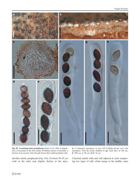
Fungal Diversity Fig. 49 Loculohypoxylon grandineum (from N.A.F. 494). a Appearance of ascomata on the host surface. b Habitat section of ascomata. c Section of an ascoma. Note the pale brown thin-walled peridium cells. slot-like ostiole, periphysate (Fig. 50a). Peridium 30–45 μm wide at the sides and slightly thicker at the apex, d, e Uniseriate ascospores in asci. f–f Cylindro-clavate asci with ascospores. Note the ocular chamber in (g). Scale bars: a=100 μm, b=200 μm, c=50 μm, d–h=10 μm 2-layered, lateral walls and wall adjacent to neck comprising two types of cells which merge in the middle; outer
Fungal Diversity cells small heavily pigmented thick-walled cells of textura angularis, cells 4–7 μm diam., cell wall 3.5–5 μm thick, inner cells less pigmented, comprising thin-walled compressed cells; apical wall cells smaller and walls thicker, basal wall thinner (ca. 15μm wide), composed of lightly pigmented thin-walled compressed cells (Fig. 50b and c). Hamathecium of trabeculate pseudoparaphyses, 1–2 μm broad, septate, anastomosing and branching rarely between and mostly above the asci. Asci 105–130(−150)×10–15 μm (x ¼ 123 12mm, n=10), 8-spored, bitunicate, fissitunicate dehiscence not observed, clavate to cylindro-clavate, with a short, narrow, furcate pedicel which is 10–25 μm long, and a small inconspicuous ocular chamber (to 1.5 μm wide× 1 μm high) (Fig. 50d and e). Ascospores (80-)90–115×3– 5 μm (x ¼ 95 3:5mm, n=10), filliform, gradually tapering towards the base, hyaline to light yellow, (6-)7(−8)-septate, slightly constricted at each septum, smooth (Fig. 50f). Anamorph: none reported. Material examined: USA, New Jersey, Newfield, on dead stems of Oenothera biennis, Aug. 1881, Ellis (NY 643, holotype, NY 885, isotype). Notes Morphology Lophionema is a relatively poorly studied genus, which was formally established by Saccardo (1883) as a monotypic genus represented by L. vermisporum based on its “globose ascomata, compressed ostiole, cylindrical to clavate ascus, and filamentous, septate, subhyaline to lightly pigmented ascospores”. Lophionema vermisporum was consequently listed as the generic type (Clements and Shear 1931). Berlese (1890) placed the genus in Lophiostomataceae but mentioned that the genus was similar to Ophiobolus according to the variable apex, and Shoemaker (1976) transferred Lophionema vermisporum to Ophiobolus sensu lato. Chesters and Bell (1970) however, had regarded Lophionema as related to Lophiostoma despite the distinct ascospore morphology. Barr (1992b) assignedLophionema to Entodesmium based on the morphology of ascomata, papilla, peridium structure, pseudoparaphyses as well as the hyaline or slightly yellowish ascospores with a terminal appendage (not observed here). Species of Entodesmium, however, exclusively occur on legumes, but Lophionema vermisporum does not. We also note that the filliform ascospores, bitunicate asci, pseudoparaphyses and nature of the peridium may also be considered as typical of genera in the Tubeufiaceae (Barr 1980; Kodsueb et al. 2006b). Phylogenetic study None. Concluding remarks The immersed to erumpent ascomata, trabeculate pseudoparaphyses and laterally flattened papilla and periphysate ostioles indicate that this genus should be included in Lophiostomataceae. We do not accept the above proposals and, consider that Lophionema should be maintained as a separate genus with filliform ascospores in Lophiostomataceae until representative taxa can be sequenced and analyzed. Currently Lophionema comprises 10 species (http://www.mycobank.org, 08-01-2009). However, many of these are poorly studied and obscure. Lophiostoma Ces. & De Not., Comm. Soc. crittog. Ital. 1: 219 (1863). (Lophiostomataceae) Generic description Habitat terrestrial, saprobic. Ascomata immersed to erumpent, usually with a distinct depressed papilla and a slotlike ostiole. Hamathecium of dense, long, septate pseudoparaphyses, embedded in mucilage, anastomosing and branching between and above the asci. Peridium unequal in thickness, thicker near the apex and thinner at base. Asci usually clavate. Ascospores 1-septate, multi-septate or even muriform, hyaline to deep brown, usually with terminal appendages. Anamorphs reported for genus: Pleuorphomopsis-like (Hyde et al. 2011). Literature: Barr 1990a; Chesters and Bell 1970; Holm and Holm 1988; Hyde and Aptroot 1998; Hyde et al. 2002; Tanaka and Harada 2003b; Yuan and Zhao 1994. Type species Lophiostoma macrostomum (Tode) Ces. & De Not., Comm. Soc. crittog. Ital. 1: 219 (1863). (Fig. 51) ≡ Sphaeria macrostoma Tode, Fung. mecklenb. sel. (Lüneburg) 2: 12 (1791). Ascomata 400–600 μm high×420–560 μm diam., densely scattered to gregarious, semi-immersed to erumpent, globose or subglobose, with a small to large flattenedcrest-likeraisedareaabovetheascomatawhich is variable in shape, up to 300 μm high and 480 μm wide, with a slit-like ostiole along the full length of the crest (Fig. 51a and b). Peridium 30–45 μm thick at the sides, thicker at the apex and thinner at the base, composed of one cell type of small lightly pigmented thin-walled cells of textura prismatica, cellsca. 6–9×3– 4 μm diam., apex composed of pseudoparenchymatous cells (Fig. 51b). Hamathecium of dense, filliform, up to 3 μm near the base and less than 1.5 μm broad in the
- Page 1 and 2:
Fungal Diversity DOI 10.1007/s13225
- Page 3 and 4:
Fungal Diversity Table 1 Major circ
- Page 5 and 6:
Fungal Diversity
- Page 7 and 8:
Fungal Diversity biocontrol agent o
- Page 9 and 10:
Fungal Diversity substrates and man
- Page 11 and 12:
Fungal Diversity 2. To investigate
- Page 13 and 14:
Fungal Diversity Table 3 (continued
- Page 15 and 16:
Fungal Diversity Table 3 (continued
- Page 17 and 18:
Fungal Diversity Table 3 (continued
- Page 19 and 20:
Fungal Diversity
- Page 21 and 22:
Fungal Diversity Fig. 2 Aigialus gr
- Page 23 and 24:
Fungal Diversity Fig. 3 Amniculicol
- Page 25 and 26:
Fungal Diversity Literature: Berkel
- Page 27 and 28:
Fungal Diversity Ascorhombispora L.
- Page 29 and 30:
Fungal Diversity
- Page 31 and 32:
Fungal Diversity Fig. 8 Astrosphaer
- Page 33 and 34:
Fungal Diversity Fig. 9 Asymmetrico
- Page 35 and 36:
Fungal Diversity Notes Morphology B
- Page 37 and 38:
Fungal Diversity Generic descriptio
- Page 39 and 40:
Fungal Diversity Anamorph: none rep
- Page 41 and 42:
Fungal Diversity Fig. 14 Bimuria no
- Page 43 and 44:
Fungal Diversity Fig. 15 Bricookea
- Page 45 and 46:
Fungal Diversity Fig. 16 Byssolophi
- Page 47 and 48:
Fungal Diversity Notes Morphology B
- Page 49 and 50: Fungal Diversity the reaction of pe
- Page 51 and 52: Fungal Diversity
- Page 53 and 54: Fungal Diversity Fig. 21 Chaetomast
- Page 55 and 56: Fungal Diversity
- Page 57 and 58: Fungal Diversity Fig. 23 Cilioplea
- Page 59 and 60: Fungal Diversity with one or two ve
- Page 61 and 62: Fungal Diversity Moreau 1953; Munk
- Page 63 and 64: Fungal Diversity Material examined:
- Page 65 and 66: Fungal Diversity Fig. 28 Dothidotth
- Page 67 and 68: Fungal Diversity Fig. 29 Dubitatio
- Page 69 and 70: Fungal Diversity assigned Entodesmi
- Page 71 and 72: Fungal Diversity fusoid to somewhat
- Page 73 and 74: Fungal Diversity Fig. 33 Hadrospora
- Page 75 and 76: Fungal Diversity Fig. 34 Halotthia
- Page 77 and 78: Fungal Diversity Notes Morphology H
- Page 79 and 80: Fungal Diversity some effused Hypox
- Page 81 and 82: Fungal Diversity Fig. 38 Isthmospor
- Page 83 and 84: Fungal Diversity Fig. 39 Kalmusia e
- Page 85 and 86: Fungal Diversity ascospores were br
- Page 87 and 88: Fungal Diversity furcate pedicel an
- Page 89 and 90: Fungal Diversity Anamorph: none rep
- Page 91 and 92: Fungal Diversity
- Page 93 and 94: Fungal Diversity Material examined:
- Page 95 and 96: Fungal Diversity Fig. 46 Lewia scro
- Page 97 and 98: Fungal Diversity Fig. 47 Lichenopyr
- Page 99: Fungal Diversity Loculohypoxylon M.
- Page 103 and 104: Fungal Diversity upper place, septa
- Page 105 and 106: Fungal Diversity
- Page 107 and 108: Fungal Diversity (CBS 627.86) was i
- Page 109 and 110: Fungal Diversity Fig. 54 Mamillisph
- Page 111 and 112: Fungal Diversity Fig. 55 Massarina
- Page 113 and 114: Fungal Diversity phaeria as a synon
- Page 115 and 116: Fungal Diversity 5-8 μm diam., ind
- Page 117 and 118: Fungal Diversity cell wall
- Page 119 and 120: Fungal Diversity Fig. 60 Mixtura sa
- Page 121 and 122: Fungal Diversity Fig. 61 Montagnula
- Page 123 and 124: Fungal Diversity spored, bitunicate
- Page 125 and 126: Fungal Diversity Fig. 64 Murispora
- Page 127 and 128: Fungal Diversity Type species Neoph
- Page 129 and 130: Fungal Diversity brown, 8-septate,
- Page 131 and 132: Fungal Diversity Fig. 68 Ohleria mo
- Page 133 and 134: Fungal Diversity Fig. 69 Ohleriella
- Page 135 and 136: Fungal Diversity Fig. 70 Ophiobolus
- Page 137 and 138: Fungal Diversity Type species Ostro
- Page 139 and 140: Fungal Diversity
- Page 141 and 142: Fungal Diversity (Shoemaker and Bab
- Page 143 and 144: Fungal Diversity ium thin, composed
- Page 145 and 146: Fungal Diversity Fig. 76 Platysporo
- Page 147 and 148: Fungal Diversity Fig. 77 1 Pleomass
- Page 149 and 150: Fungal Diversity Fig. 78 Pleophragm
- Page 151 and 152:
Fungal Diversity papillate, ostiola
- Page 153 and 154:
Fungal Diversity Williams 1963; Mal
- Page 155 and 156:
Fungal Diversity Generic descriptio
- Page 157 and 158:
Fungal Diversity composed of one ce
- Page 159 and 160:
Fungal Diversity Fig. 84 Saccharico
- Page 161 and 162:
Fungal Diversity and nearly black a
- Page 163 and 164:
Fungal Diversity dense, long trabec
- Page 165 and 166:
Fungal Diversity
- Page 167 and 168:
Fungal Diversity
- Page 169 and 170:
Fungal Diversity Anamorphs reported
- Page 171 and 172:
Fungal Diversity
- Page 173 and 174:
Fungal Diversity
- Page 175 and 176:
Fungal Diversity Fig. 94 Westerdyke
- Page 177 and 178:
Fungal Diversity Fig. 95 Wettsteini
- Page 179 and 180:
Fungal Diversity Fig. 96 Wilmia bra
- Page 181 and 182:
Fungal Diversity Current name: Astr
- Page 183 and 184:
Fungal Diversity spores are actuall
- Page 185 and 186:
Fungal Diversity Fig. 100 Sporormie
- Page 187 and 188:
Fungal Diversity
- Page 189 and 190:
Fungal Diversity Fig. 102 Kriegerie
- Page 191 and 192:
Fungal Diversity Phylogenetic study
- Page 193 and 194:
Fungal Diversity Fig. 104 Zeuctomor
- Page 195 and 196:
Fungal Diversity Fig. 105 Muroia ni
- Page 197 and 198:
Fungal Diversity pseudoparenchymato
- Page 199 and 200:
Fungal Diversity Eremodothis Arx, K
- Page 201 and 202:
Fungal Diversity Type species: Macr
- Page 203 and 204:
Fungal Diversity ascospores of Plat
- Page 205 and 206:
Fungal Diversity monoceras Alcorn n
- Page 207 and 208:
Fungal Diversity tomataceae, Melano
- Page 209 and 210:
Fungal Diversity Table 4 (continued
- Page 211 and 212:
Fungal Diversity 1987b). Based on a
- Page 213 and 214:
Fungal Diversity only do so under v
- Page 215 and 216:
Fungal Diversity Dennis RWG (1968)
- Page 217 and 218:
Fungal Diversity Kirk PM, Cannon PF
- Page 219 and 220:
Fungal Diversity Saccardo PA (1880)
- Page 221:
Fungal Diversity Winter G (1887) As