

Pleosporales - CBS - KNAW
Pleosporales - CBS - KNAW
Pleosporales - CBS - KNAW

Create successful ePaper yourself
Turn your PDF publications into a flip-book with our unique Google optimized e-Paper software.
Fungal Diversity<br />
Anamorph: Pycnidia typical of Stagonospora (Sphaeropsidales),<br />
“scattered,arisingsinglybothonthehostandinpure<br />
culture, in culture generally surrounded by an envelope of<br />
mycelial hyphae, numerous, immersed on the host, but nearly<br />
superficial in culture, subglobose to slightly applanate, black,<br />
150–250 μm diam., with a central slightly papillate ostiole,<br />
lacking a distinct neck; walls mainly 15–20 μm thick,<br />
composed of three to six layers of pseudoparenchymatous<br />
cells, the outermost layers dark brown and inner pale brown to<br />
hyaline cells somewhat compressed radially, very variable in<br />
size, cells of the outer layers mainly 7–12 μm long×4–6 μm<br />
wide in vertically section and 10–12 μm diam. in surface<br />
view, wall not or only slightly thicked near the ostiole.<br />
Conidiogenous cells lining the inner surface of the pycnidial<br />
cavity, holoblastic, minute and difficult to distinguish from the<br />
pseudoparenchymatous cells with which they are mixed,<br />
mammiform with a flattened apex, hyaline, smooth walled,<br />
about 4–6 μm tall and 4–6 μm wide. Conidia copiously<br />
produced, ellipsoid, with somewhat truncated ends, hyaline,<br />
smooth walled, (2-)3 septate, not or slightly constricted at the<br />
septa, often guttulate, rather thin walled, (21-)24–28(−34)<br />
μm×7–8.5(−11.5) μm” (from Kaiser et al. 1979).<br />
Material examined: KENYA, near Nairobi, on leaves of<br />
Saccharum officinarum L.; 24 Aug. 1977; leg. W.J. Kaiser<br />
(IMI 215888, holotype).<br />
Notes<br />
Morphology<br />
Saccharicola was separated from Leptosphaeria as a new<br />
genus based on its Stagonospora anamorph and its biotrophic<br />
habitat in leaves of sugar cane, and two species were<br />
included, i.e. Saccharicola bicolor and S. taiwanensis (J.M.<br />
Yen & C.C. Chi) O.E. Erikss. & D. Hawksw. (Eriksson and<br />
Hawksworth 2003). Saccharicola is characterized by its<br />
parasitic habitat on monocots, small ascomata, bitunicate<br />
asci, presence of pseudoparaphyses as well as its 3-septate<br />
ascospores (Eriksson and Hawksworth 2003).<br />
Phylogenetic study<br />
Based on the limited phylogenetic analysis of SSU<br />
sequences, Saccharicola is considered to be closely related<br />
to Massarina eburnea, the generic type of Massarina<br />
(Eriksson and Hawksworth 2003). Thus, Saccharicola was<br />
assigned to Massarinaceae, which includes Keissleriella,<br />
Massarina and Saccharicola (Eriksson and Hawksworth<br />
2003).<br />
Concluding remarks<br />
Based on the parasitic habitat on monocots and its small<br />
ascomata and Stagonospora (or Cercospora? for S. taiwanensis,<br />
see Eriksson and Hawksworth 2003; Shoemaker<br />
and Babcock 1989b) anamorph, Saccharicola seems more<br />
similar to Pleosporineae. Further molecular study is needed<br />
for confirmation.<br />
Salsuginea K.D. Hyde, Bot. Mar. 34: 315 (1991). (<strong>Pleosporales</strong>,<br />
genera incertae sedis)<br />
Generic description<br />
Habitat marine, saprobic. Ascomata large, solitary, fusoid,<br />
conical or subglobose, with or without a flattened base,<br />
immersed under a darkened clypeus, papillate, ostiolate.<br />
Peridium thin, composed of round cells (in cross section) at<br />
sides, fusing at the top with the clypeus, thin at the base.<br />
Hamathecium of dense, long trabeculate pseudoparaphyses,<br />
anastomosing, embedded in mucilage. Asci 8-spored, bitunicate,<br />
fissitunicate, clavate to cylindro-clavate, pedunculate,<br />
with a large ocular chamber and conspicuous apical ring.<br />
Ascospores uniseriate, obovoid, brown to black, with hyaline<br />
apical germ pores, 1-septate, constricted at the septum, dark<br />
brown with paler apical cells, lacking sheath, smooth.<br />
Anamorphs reported for genus: none.<br />
Literature: Hyde 1991a; Suetrong et al. 2009.<br />
Type species<br />
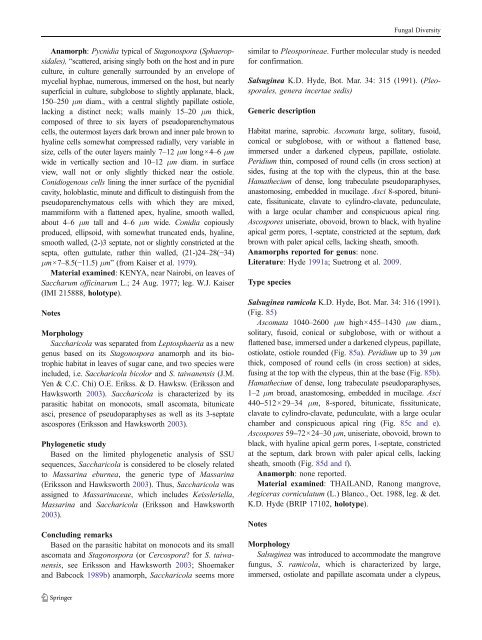
Salsuginea ramicola K.D. Hyde, Bot. Mar. 34: 316 (1991).<br />
(Fig. 85)<br />
Ascomata 1040–2600 μm high×455–1430 μm diam.,<br />
solitary, fusoid, conical or subglobose, with or without a<br />
flattened base, immersed under a darkened clypeus, papillate,<br />
ostiolate, ostiole rounded (Fig. 85a). Peridium up to 39 μm<br />
thick, composed of round cells (in cross section) at sides,<br />
fusing at the top with the clypeus, thin at the base (Fig. 85b).<br />
Hamathecium of dense, long trabeculate pseudoparaphyses,<br />
1–2 μm broad, anastomosing, embedded in mucilage. Asci<br />
440–512×29–34 μm, 8-spored, bitunicate, fissitunicate,<br />
clavate to cylindro-clavate, pedunculate, with a large ocular<br />
chamber and conspicuous apical ring (Fig. 85c and e).<br />
Ascospores 59–72×24–30 μm, uniseriate, obovoid, brown to<br />
black, with hyaline apical germ pores, 1-septate, constricted<br />
at the septum, dark brown with paler apical cells, lacking<br />
sheath, smooth (Fig. 85d and f).<br />
Anamorph: none reported.<br />
Material examined: THAILAND, Ranong mangrove,<br />
Aegiceras corniculatum (L.) Blanco., Oct. 1988, leg. & det.<br />
K.D. Hyde (BRIP 17102, holotype).<br />
Notes<br />
Morphology<br />
Salsuginea was introduced to accommodate the mangrove<br />
fungus, S. ramicola, which is characterized by large,<br />
immersed, ostiolate and papillate ascomata under a clypeus,


