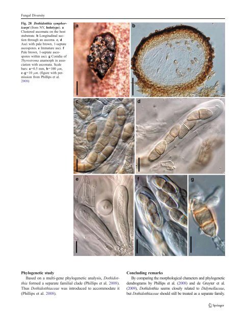
Fungal Diversity 1988; Scheinpflug 1958). Over 400 epithets of Didymosphaeria were included until the monograph of Aptroot (1995). Aptroot (1995) examined more than 3000 specimens under the name Didymosphaeria. The type specimen of Didymosphaeria (Fungi Rhenani 1770) represents the widespread and common D. futilis (Aptroot 1995). In this study, we did not get the lectotype specimen, but described the type of D. futilis (Sphaeria futilis). Using a narrow concept (ignoring differences of host or country of origin), Aptroot (1995) accepted only seven species, which were closely related with the generic type of Didymosphaeria with over 100 synonyms distributed among them. Many taxa were found to belong to other groups, i.e. Aaosphaeria, Amphisphaeria, Astrosphaeriella, Dothidotthia, Flagellosphaeria, Kirschsteiniothelia, Megalotremis, Montagnula, Munkovalsaria, Mycomicrothelia, Parapyrenis or Phaeodothis. Didymosphaeria is mainly characterized by a peridium consisting of flattened or irregular cells or completely hyphae; a hamathecium consisting of narrow, trabeculate paraphysoids or paraphyses, richly anastomosing above the asci; and brown thinly distoseptate ascospores. Didymosphaeriaceae was maintained as a separated family within <strong>Pleosporales</strong> by Aptroot (1995) because of the distoseptate ascospores and trabeculate pseudoparaphyses mainly anastomosing above the asci. This proposal, however, has not received much support (Lumbsch and Huhndorf 2007). Phylogenetic study There have been few molecular investigations of Didymosphaeria when compared to the morphological studies. Didymosphaeria futilis resided in the clade of Cucurbitariaceae (or Didymosphaeriaceae) (Plate 1). The correct identification of the Didymosphaeria strain used for sequencing, however, has not been verified. Concluding remarks Didymosphaeria is a well established genus represented by D. futilis. Of particular significance are the narrow pseudoparaphyses which anastomose above the asci and brown 1-septate ascospores with indistinct distosepta. Familial placement of Didymosphaeria is unclear yet because of insufficient molecular data. Dothidotthia Höhn., Ber. Deutsch. Bot. Ges. 36: 312 (1918). (Didymellaceae) Generic description Habitat terrestrial, saprobic. Ascomata medium-sized, solitary, clustered or somewhat gregarious, erumpent, subglobose, apex somewhat papillate to depressed, coriaceous. Peridium composed of a few layers of dark brown cells of textura angularis, and giving rise dark brown, thick-walled hyphae in the basal region, 2-layered. Hamathecium septate pseudoparaphyses branched in upper part above asci. Asci 8-spored, bitunicate, clavate, straight to curved. Ascospores biseriate to obliquely uniseriate, ellipsoid, pale brown, 1-septate. Anamorphs reported for genus: Dothiorella and Thyrostroma (Hyde et al. 2011; Phillips et al. 2008). Literature: Barr1989b; Phillips et al. 2008. Type species Dothidotthia symphoricarpi (Rehm) Höhn., Ber. Deutsch. Bot. Ges. 36: 312 (1918). (Fig. 28) ≡ Pseudotthia symphoricarpi Rehm, Ann. Mycol. 11: 169 (1913). Ascomata up to 500 μm high×550 μm diam., gregarious clustered, rarely solitary, erumpent, subglobose, apex somewhat papillate to depressed, coriaceous (Fig. 28a). Peridium 20– 80 μm thick, composed of 3–6 layers of dark brown cells of textura angularis, giving rise dark brown, thick-walled hyphae in the basal region, 2-layered, outer layer wall thicker and inner layer wall thinner (Fig. 28b). Hamathecium hyaline, septate pseudoparaphyses, 2–3 μm wide, branched in upper part above asci. Asci 70–120×15–22 μm, 8-spored, bitunicate, clavate, straight to curved (Fig. 28c, d and e). Ascospores (20-)22–23 (−26)×(8-)9–10(−11) μm, biseriate to obliquely uniseriate and partially overlapping, ellipsoid tapering towards subacutely rounded ends, pale brown, 1-septate, constricted at the septum, smooth (Fig. 28f) (description referred to Phillips et al. 2008). Anamorph: Thyrostroma negundinis (Phillips et al. 2008). Material examined: USA, North Dakota, on branches of Symphoricarpos occidentalis Hook. (NY, holotype); Colorado, San Juan Co, c. 0.5 mile up Engineer Mountain Trail from turnoff at mile 52.5, Hwy 550, dead twigs of Symphoricarpos rotundifolius A. Gray, 24 Jun. 2004, A.W. Ramaley 0410 (BPI 871823, epitype). Notes Morphology Dothidotthia was formally established to accommodate Pseudotthia symphoricarpi (Montagnellaceae, Dothideales) (von Höhnel 1918a). Many mycologists considered Dothidotthia closely related to a genus of Venturiaceae such as Dibotryon by Petrak (1927), or Gibbera by von Arx and Müller (1954) and Müller and von Arx (1962). Dothidotthia had been treated as a synonym of Gibbera (von Arx 1954; Müller and von Arx 1962), which was followed by Shoemaker (1963) and Eriksson and Hawksworth (1987). Based on the coelomycetous anamorphic stage and peridium structure, shape of asci, as well as morphology of pseudoparaphyses, Barr (1987b, 1989b) retrieved Dothidotthia, and considered it closely related to Botryosphaeria (Botryosphaeriaceae). Currently, 11 species are included within Dothidotthia (http://www.mycobank.org, 01–2011).
Fungal Diversity Fig. 28 Dothidotthia symphoricarpi (from NY, holotype). a Clustered ascomata on the host stubstrate. b Longitudinal section through an ascoma. c, d Asci with pale brown, 1-septate ascospores. e Immature asci. f Pale brown, 1-septate ascospores within asci. g Conidia of Thyrostroma anamorph in association with ascomata. Scale bars: a=0.5 mm, b=100 μm, c–g=10 μm. (figure with permission from Phillips et al. 2008) Phylogenetic study Based on a multi-gene phylogenetic analysis, Dothidotthia formed a separate familial clade (Phillips et al. 2008). Thus Dothidotthiaceae was introduced to accommodate it (Phillips et al. 2008). Concluding remarks By comparing the morphological characters and phylogenetic dendrograms by Phillips et al. (2008) and de Gruyter et al. (2009), Dothidotthia seems closely related to Didymellaceae, but Dothidotthiaceae should still be treated as a separate family.
- Page 1 and 2:
Fungal Diversity DOI 10.1007/s13225
- Page 3 and 4:
Fungal Diversity Table 1 Major circ
- Page 5 and 6:
Fungal Diversity
- Page 7 and 8:
Fungal Diversity biocontrol agent o
- Page 9 and 10:
Fungal Diversity substrates and man
- Page 11 and 12:
Fungal Diversity 2. To investigate
- Page 13 and 14: Fungal Diversity Table 3 (continued
- Page 15 and 16: Fungal Diversity Table 3 (continued
- Page 17 and 18: Fungal Diversity Table 3 (continued
- Page 19 and 20: Fungal Diversity
- Page 21 and 22: Fungal Diversity Fig. 2 Aigialus gr
- Page 23 and 24: Fungal Diversity Fig. 3 Amniculicol
- Page 25 and 26: Fungal Diversity Literature: Berkel
- Page 27 and 28: Fungal Diversity Ascorhombispora L.
- Page 29 and 30: Fungal Diversity
- Page 31 and 32: Fungal Diversity Fig. 8 Astrosphaer
- Page 33 and 34: Fungal Diversity Fig. 9 Asymmetrico
- Page 35 and 36: Fungal Diversity Notes Morphology B
- Page 37 and 38: Fungal Diversity Generic descriptio
- Page 39 and 40: Fungal Diversity Anamorph: none rep
- Page 41 and 42: Fungal Diversity Fig. 14 Bimuria no
- Page 43 and 44: Fungal Diversity Fig. 15 Bricookea
- Page 45 and 46: Fungal Diversity Fig. 16 Byssolophi
- Page 47 and 48: Fungal Diversity Notes Morphology B
- Page 49 and 50: Fungal Diversity the reaction of pe
- Page 51 and 52: Fungal Diversity
- Page 53 and 54: Fungal Diversity Fig. 21 Chaetomast
- Page 55 and 56: Fungal Diversity
- Page 57 and 58: Fungal Diversity Fig. 23 Cilioplea
- Page 59 and 60: Fungal Diversity with one or two ve
- Page 61 and 62: Fungal Diversity Moreau 1953; Munk
- Page 63: Fungal Diversity Material examined:
- Page 67 and 68: Fungal Diversity Fig. 29 Dubitatio
- Page 69 and 70: Fungal Diversity assigned Entodesmi
- Page 71 and 72: Fungal Diversity fusoid to somewhat
- Page 73 and 74: Fungal Diversity Fig. 33 Hadrospora
- Page 75 and 76: Fungal Diversity Fig. 34 Halotthia
- Page 77 and 78: Fungal Diversity Notes Morphology H
- Page 79 and 80: Fungal Diversity some effused Hypox
- Page 81 and 82: Fungal Diversity Fig. 38 Isthmospor
- Page 83 and 84: Fungal Diversity Fig. 39 Kalmusia e
- Page 85 and 86: Fungal Diversity ascospores were br
- Page 87 and 88: Fungal Diversity furcate pedicel an
- Page 89 and 90: Fungal Diversity Anamorph: none rep
- Page 91 and 92: Fungal Diversity
- Page 93 and 94: Fungal Diversity Material examined:
- Page 95 and 96: Fungal Diversity Fig. 46 Lewia scro
- Page 97 and 98: Fungal Diversity Fig. 47 Lichenopyr
- Page 99 and 100: Fungal Diversity Loculohypoxylon M.
- Page 101 and 102: Fungal Diversity cells small heavil
- Page 103 and 104: Fungal Diversity upper place, septa
- Page 105 and 106: Fungal Diversity
- Page 107 and 108: Fungal Diversity (CBS 627.86) was i
- Page 109 and 110: Fungal Diversity Fig. 54 Mamillisph
- Page 111 and 112: Fungal Diversity Fig. 55 Massarina
- Page 113 and 114: Fungal Diversity phaeria as a synon
- Page 115 and 116:
Fungal Diversity 5-8 μm diam., ind
- Page 117 and 118:
Fungal Diversity cell wall
- Page 119 and 120:
Fungal Diversity Fig. 60 Mixtura sa
- Page 121 and 122:
Fungal Diversity Fig. 61 Montagnula
- Page 123 and 124:
Fungal Diversity spored, bitunicate
- Page 125 and 126:
Fungal Diversity Fig. 64 Murispora
- Page 127 and 128:
Fungal Diversity Type species Neoph
- Page 129 and 130:
Fungal Diversity brown, 8-septate,
- Page 131 and 132:
Fungal Diversity Fig. 68 Ohleria mo
- Page 133 and 134:
Fungal Diversity Fig. 69 Ohleriella
- Page 135 and 136:
Fungal Diversity Fig. 70 Ophiobolus
- Page 137 and 138:
Fungal Diversity Type species Ostro
- Page 139 and 140:
Fungal Diversity
- Page 141 and 142:
Fungal Diversity (Shoemaker and Bab
- Page 143 and 144:
Fungal Diversity ium thin, composed
- Page 145 and 146:
Fungal Diversity Fig. 76 Platysporo
- Page 147 and 148:
Fungal Diversity Fig. 77 1 Pleomass
- Page 149 and 150:
Fungal Diversity Fig. 78 Pleophragm
- Page 151 and 152:
Fungal Diversity papillate, ostiola
- Page 153 and 154:
Fungal Diversity Williams 1963; Mal
- Page 155 and 156:
Fungal Diversity Generic descriptio
- Page 157 and 158:
Fungal Diversity composed of one ce
- Page 159 and 160:
Fungal Diversity Fig. 84 Saccharico
- Page 161 and 162:
Fungal Diversity and nearly black a
- Page 163 and 164:
Fungal Diversity dense, long trabec
- Page 165 and 166:
Fungal Diversity
- Page 167 and 168:
Fungal Diversity
- Page 169 and 170:
Fungal Diversity Anamorphs reported
- Page 171 and 172:
Fungal Diversity
- Page 173 and 174:
Fungal Diversity
- Page 175 and 176:
Fungal Diversity Fig. 94 Westerdyke
- Page 177 and 178:
Fungal Diversity Fig. 95 Wettsteini
- Page 179 and 180:
Fungal Diversity Fig. 96 Wilmia bra
- Page 181 and 182:
Fungal Diversity Current name: Astr
- Page 183 and 184:
Fungal Diversity spores are actuall
- Page 185 and 186:
Fungal Diversity Fig. 100 Sporormie
- Page 187 and 188:
Fungal Diversity
- Page 189 and 190:
Fungal Diversity Fig. 102 Kriegerie
- Page 191 and 192:
Fungal Diversity Phylogenetic study
- Page 193 and 194:
Fungal Diversity Fig. 104 Zeuctomor
- Page 195 and 196:
Fungal Diversity Fig. 105 Muroia ni
- Page 197 and 198:
Fungal Diversity pseudoparenchymato
- Page 199 and 200:
Fungal Diversity Eremodothis Arx, K
- Page 201 and 202:
Fungal Diversity Type species: Macr
- Page 203 and 204:
Fungal Diversity ascospores of Plat
- Page 205 and 206:
Fungal Diversity monoceras Alcorn n
- Page 207 and 208:
Fungal Diversity tomataceae, Melano
- Page 209 and 210:
Fungal Diversity Table 4 (continued
- Page 211 and 212:
Fungal Diversity 1987b). Based on a
- Page 213 and 214:
Fungal Diversity only do so under v
- Page 215 and 216:
Fungal Diversity Dennis RWG (1968)
- Page 217 and 218:
Fungal Diversity Kirk PM, Cannon PF
- Page 219 and 220:
Fungal Diversity Saccardo PA (1880)
- Page 221:
Fungal Diversity Winter G (1887) As