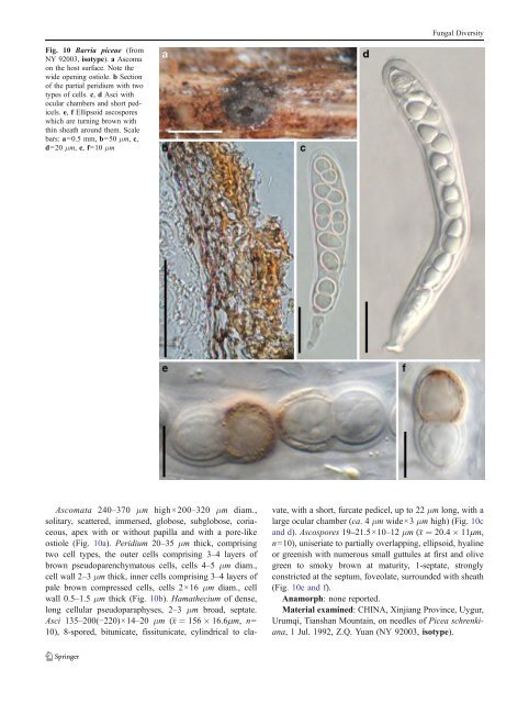
Fungal Diversity Fig. 10 Barria piceae (from NY 92003, isotype). a Ascoma on the host surface. Note the wide opening ostiole. b Section of the partial peridium with two types of cells. c, d Asci with ocular chambers and short pedicels. e, f Ellipsoid ascospores which are turning brown with thin sheath around them. Scale bars: a=0.5 mm, b=50 μm, c, d=20 μm, e, f=10 μm Ascomata 240–370 μm high×200–320 μm diam., solitary, scattered, immersed, globose, subglobose, coriaceous, apex with or without papilla and with a pore-like ostiole (Fig. 10a). Peridium 20–35 μm thick, comprising two cell types, the outer cells comprising 3–4 layers of brown pseudoparenchymatous cells, cells 4–5 μm diam., cell wall 2–3 μm thick, inner cells comprising 3–4 layers of pale brown compressed cells, cells 2×16 μm diam., cell wall 0.5–1.5 μm thick (Fig. 10b). Hamathecium of dense, long cellular pseudoparaphyses, 2–3 μm broad, septate. Asci 135–200(−220)×14–20 μm (x ¼ 156 16:6mm, n= 10), 8-spored, bitunicate, fissitunicate, cylindrical to clavate, with a short, furcate pedicel, up to 22 μm long, with a large ocular chamber (ca. 4μm wide×3 μm high) (Fig. 10c and d). Ascospores 19–21.5×10⁻¹² μm (x ¼ 20:4 11mm, n=10), uniseriate to partially overlapping, ellipsoid, hyaline or greenish with numerous small guttules at first and olive green to smoky brown at maturity, 1-septate, strongly constricted at the septum, foveolate, surrounded with sheath (Fig. 10e and f). Anamorph: none reported. Material examined: CHINA, Xinjiang Province, Uygur, Urumqi, Tianshan Mountain, on needles of Picea schrenkiana, 1 Jul. 1992, Z.Q. Yuan (NY 92003, isotype).
Fungal Diversity Notes Morphology Barria was established by Yuan (1994) as a monotypic genus represented by B. piceae according to its “two-celled, pigmented ascospores, pseudoparenchymatous peridium and narrowly cellular pseudoparaphyses” thus differing in its combination of characters from all of the morphologically related dothideomycetous genera, such as Didymosphaeria, Didymopleella or Stegasphaeria. The taxon was considered to belong in Phaeosphaeriaceae. Ascomata and colour or shape of ascospores, however, readily distinguish it from other 1-septate Phaeosphaeriaceae genera, i.e. Didymella, Lautitia and Metameris (Yuan 1994). Barria piceae causes blight of spruce needles. Phylogenetic study None. Concluding remarks The status of Barria with its unusual verrucose ascospores and thick gel coating is uncertain. In many ways it resembles Belizeana, with its cylindrical asci, 1-septate, ellipsoid ascospores with sheath and verruculose surface (Kohlmeyer and Volkmann-Kohlmeyer 1987). However, the latter is a marine genus while Barria causes leaf blight of terrestrial Picea (Yuan 1994). The placement in Phaeosphaeriaceae seems logical based on the parasitic life style, thin and simple peridium, wide cellular pseudoparaphyses and brown ascospores. However, molecular data are needed to confirm this. Belizeana Kohlm. & Volkm.-Kohlm., Bot. Mar. 30: 195 (1987). (<strong>Pleosporales</strong>, genera incertae sedis) Generic description Habitat marine, saprobic. Ascomata solitary, scattered, or in small groups, medium-sized, immersed to semiimmersed, subglobose to broadly ampulliform, black, ostiolate, carbonaceous. Peridium thin, comprising several layers of brown thin-walled cells of textura angularis. Hamathecium of dense, filliform pseudoparaphyses, rarely branched. Asci 8-spored, bitunicate, fissitunicate, broadly cylindrical to clavate, with a short pedicel and an ocular chamber. Ascospores uniseriate, broadly ellipsoidal, hyaline, turn pale brown when senescent, 1-septate, constricted at the septum, thick-walled, 2-layered, mature spores with tuberculate ornamentation between the two layers. Anamorphs reported for genus: Phoma-like (Kohlmeyer and Volkmann-Kohlmeyer 1987). Literature: Kohlmeyer and Volkmann-Kohlmeyer 1987. Type species Belizeana tuberculata Kohlm. & Volkm.-Kohlm., Bot. Mar. 30: 196 (1987). (Fig. 11) Ascomata 170–300 μm high×160–290 μm diam., solitary, scattered, or in small groups of 2–3, immersed to semiimmersed, subglobose to broadly ampulliform, carbonaceous, black, pale brown on the sides, ostiolate, epapillate or shortly papillate, ostiolar canal filled with a tissue of hyaline cells (Fig. 11a). Peridium 25–35 μm wide, comprising several layers thin-walled cells of textura angularis, which are hyaline inwardly, near the base composed of a hyaline hyphal mass producing asci, up to 20 μm thick (Fig. 11b, c and e). Hamathecium of dense, ca. 2 μm broad, filliform pseudoparaphyses, rarely branched, embedded in mucilage (Fig. 11g). Asci 145–170×20–30 μm (x ¼ 163 25mm, n= 10), 8-spored, bitunicate, fissitunicate, broadly cylindrical to clavate with a short pedicel, thick-walled, with a small ocular chamber (Fig. 11d, f and h). Ascospores 21–26 × 13–18 μm (x ¼ 22 15mm, n = 10), uniseriate, broadly ellipsoidal, hyaline, turn pale brown when senescent, 1-septate, constricted at the septum, thick-walled, 2-layered, mature spores with tuberculate ornamentation between the two layers (Fig. 11i and j). Anamorph: Phoma-like (Kohlmeyer and Volkmann- Kohlmeyer 1987). Material examined: BELIZE, Twin Cays, on Laguncularia sp., 7 Apr. 1983, leg. & det. J. Kohlmeyer (Herb. J. Kohlmeyer No. 4398, holotype); AUSTRALIA, Towra Point, New South Wales, trunk of eroded tree with oysters and shipworms, intertidal zone, Botany Bay, 23 Aug. 1981 (Herb. J. Kohlmeyer No. 4209, paratype). Notes Morphology Belizeana was formally established to accommodate B. tuberculata, an obligate marine fungus, which is characterized by verrucose ascospores (Kohlmeyer and Volkmann- Kohlmeyer 1987). Belizeana tuberculata canbeassignedto Pleosporaceae (<strong>Pleosporales</strong>) according to Luttrell’s (1973) treatment and keys of von Arx and Müller (1975), but cannot resolve a proper family based on Barr (1979a, 1983). The unique morphology together with obligate marine habitat makes B. tuberculata readily distinguishable from all other taxa of Pleosporaceae. Phylogenetic study None. Concluding remarks The ascospores of Belizeana tuberculata are most comparable with those of Acrocordiopsis patilii, but the superficial
- Page 1 and 2: Fungal Diversity DOI 10.1007/s13225
- Page 3 and 4: Fungal Diversity Table 1 Major circ
- Page 5 and 6: Fungal Diversity
- Page 7 and 8: Fungal Diversity biocontrol agent o
- Page 9 and 10: Fungal Diversity substrates and man
- Page 11 and 12: Fungal Diversity 2. To investigate
- Page 13 and 14: Fungal Diversity Table 3 (continued
- Page 15 and 16: Fungal Diversity Table 3 (continued
- Page 17 and 18: Fungal Diversity Table 3 (continued
- Page 19 and 20: Fungal Diversity
- Page 21 and 22: Fungal Diversity Fig. 2 Aigialus gr
- Page 23 and 24: Fungal Diversity Fig. 3 Amniculicol
- Page 25 and 26: Fungal Diversity Literature: Berkel
- Page 27 and 28: Fungal Diversity Ascorhombispora L.
- Page 29 and 30: Fungal Diversity
- Page 31 and 32: Fungal Diversity Fig. 8 Astrosphaer
- Page 33: Fungal Diversity Fig. 9 Asymmetrico
- Page 37 and 38: Fungal Diversity Generic descriptio
- Page 39 and 40: Fungal Diversity Anamorph: none rep
- Page 41 and 42: Fungal Diversity Fig. 14 Bimuria no
- Page 43 and 44: Fungal Diversity Fig. 15 Bricookea
- Page 45 and 46: Fungal Diversity Fig. 16 Byssolophi
- Page 47 and 48: Fungal Diversity Notes Morphology B
- Page 49 and 50: Fungal Diversity the reaction of pe
- Page 51 and 52: Fungal Diversity
- Page 53 and 54: Fungal Diversity Fig. 21 Chaetomast
- Page 55 and 56: Fungal Diversity
- Page 57 and 58: Fungal Diversity Fig. 23 Cilioplea
- Page 59 and 60: Fungal Diversity with one or two ve
- Page 61 and 62: Fungal Diversity Moreau 1953; Munk
- Page 63 and 64: Fungal Diversity Material examined:
- Page 65 and 66: Fungal Diversity Fig. 28 Dothidotth
- Page 67 and 68: Fungal Diversity Fig. 29 Dubitatio
- Page 69 and 70: Fungal Diversity assigned Entodesmi
- Page 71 and 72: Fungal Diversity fusoid to somewhat
- Page 73 and 74: Fungal Diversity Fig. 33 Hadrospora
- Page 75 and 76: Fungal Diversity Fig. 34 Halotthia
- Page 77 and 78: Fungal Diversity Notes Morphology H
- Page 79 and 80: Fungal Diversity some effused Hypox
- Page 81 and 82: Fungal Diversity Fig. 38 Isthmospor
- Page 83 and 84: Fungal Diversity Fig. 39 Kalmusia e
- Page 85 and 86:
Fungal Diversity ascospores were br
- Page 87 and 88:
Fungal Diversity furcate pedicel an
- Page 89 and 90:
Fungal Diversity Anamorph: none rep
- Page 91 and 92:
Fungal Diversity
- Page 93 and 94:
Fungal Diversity Material examined:
- Page 95 and 96:
Fungal Diversity Fig. 46 Lewia scro
- Page 97 and 98:
Fungal Diversity Fig. 47 Lichenopyr
- Page 99 and 100:
Fungal Diversity Loculohypoxylon M.
- Page 101 and 102:
Fungal Diversity cells small heavil
- Page 103 and 104:
Fungal Diversity upper place, septa
- Page 105 and 106:
Fungal Diversity
- Page 107 and 108:
Fungal Diversity (CBS 627.86) was i
- Page 109 and 110:
Fungal Diversity Fig. 54 Mamillisph
- Page 111 and 112:
Fungal Diversity Fig. 55 Massarina
- Page 113 and 114:
Fungal Diversity phaeria as a synon
- Page 115 and 116:
Fungal Diversity 5-8 μm diam., ind
- Page 117 and 118:
Fungal Diversity cell wall
- Page 119 and 120:
Fungal Diversity Fig. 60 Mixtura sa
- Page 121 and 122:
Fungal Diversity Fig. 61 Montagnula
- Page 123 and 124:
Fungal Diversity spored, bitunicate
- Page 125 and 126:
Fungal Diversity Fig. 64 Murispora
- Page 127 and 128:
Fungal Diversity Type species Neoph
- Page 129 and 130:
Fungal Diversity brown, 8-septate,
- Page 131 and 132:
Fungal Diversity Fig. 68 Ohleria mo
- Page 133 and 134:
Fungal Diversity Fig. 69 Ohleriella
- Page 135 and 136:
Fungal Diversity Fig. 70 Ophiobolus
- Page 137 and 138:
Fungal Diversity Type species Ostro
- Page 139 and 140:
Fungal Diversity
- Page 141 and 142:
Fungal Diversity (Shoemaker and Bab
- Page 143 and 144:
Fungal Diversity ium thin, composed
- Page 145 and 146:
Fungal Diversity Fig. 76 Platysporo
- Page 147 and 148:
Fungal Diversity Fig. 77 1 Pleomass
- Page 149 and 150:
Fungal Diversity Fig. 78 Pleophragm
- Page 151 and 152:
Fungal Diversity papillate, ostiola
- Page 153 and 154:
Fungal Diversity Williams 1963; Mal
- Page 155 and 156:
Fungal Diversity Generic descriptio
- Page 157 and 158:
Fungal Diversity composed of one ce
- Page 159 and 160:
Fungal Diversity Fig. 84 Saccharico
- Page 161 and 162:
Fungal Diversity and nearly black a
- Page 163 and 164:
Fungal Diversity dense, long trabec
- Page 165 and 166:
Fungal Diversity
- Page 167 and 168:
Fungal Diversity
- Page 169 and 170:
Fungal Diversity Anamorphs reported
- Page 171 and 172:
Fungal Diversity
- Page 173 and 174:
Fungal Diversity
- Page 175 and 176:
Fungal Diversity Fig. 94 Westerdyke
- Page 177 and 178:
Fungal Diversity Fig. 95 Wettsteini
- Page 179 and 180:
Fungal Diversity Fig. 96 Wilmia bra
- Page 181 and 182:
Fungal Diversity Current name: Astr
- Page 183 and 184:
Fungal Diversity spores are actuall
- Page 185 and 186:
Fungal Diversity Fig. 100 Sporormie
- Page 187 and 188:
Fungal Diversity
- Page 189 and 190:
Fungal Diversity Fig. 102 Kriegerie
- Page 191 and 192:
Fungal Diversity Phylogenetic study
- Page 193 and 194:
Fungal Diversity Fig. 104 Zeuctomor
- Page 195 and 196:
Fungal Diversity Fig. 105 Muroia ni
- Page 197 and 198:
Fungal Diversity pseudoparenchymato
- Page 199 and 200:
Fungal Diversity Eremodothis Arx, K
- Page 201 and 202:
Fungal Diversity Type species: Macr
- Page 203 and 204:
Fungal Diversity ascospores of Plat
- Page 205 and 206:
Fungal Diversity monoceras Alcorn n
- Page 207 and 208:
Fungal Diversity tomataceae, Melano
- Page 209 and 210:
Fungal Diversity Table 4 (continued
- Page 211 and 212:
Fungal Diversity 1987b). Based on a
- Page 213 and 214:
Fungal Diversity only do so under v
- Page 215 and 216:
Fungal Diversity Dennis RWG (1968)
- Page 217 and 218:
Fungal Diversity Kirk PM, Cannon PF
- Page 219 and 220:
Fungal Diversity Saccardo PA (1880)
- Page 221:
Fungal Diversity Winter G (1887) As