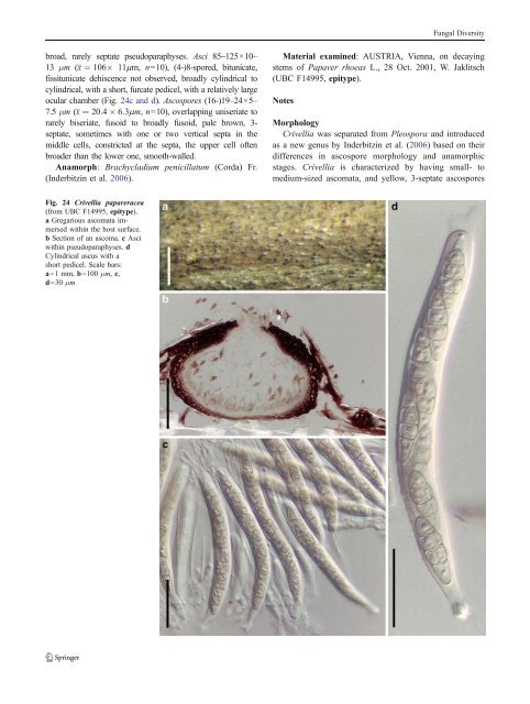
Fungal Diversity broad, rarely septate pseudoparaphyses. Asci 85–125×10– 13 μm (x ¼ 106 11mm, n=10), (4-)8-spored, bitunicate, fissitunicate dehiscence not observed, broadly cylindrical to cylindrical, with a short, furcate pedicel, with a relatively large ocular chamber (Fig. 24c and d). Ascospores (16-)19–24×5– 7.5 μm (x ¼ 20:4 6:3mm, n=10), overlapping uniseriate to rarely biseriate, fusoid to broadly fusoid, pale brown, 3- septate, sometimes with one or two vertical septa in the middle cells, constricted at the septa, the upper cell often broader than the lower one, smooth-walled. Anamorph: Brachycladium penicillatum (Corda) Fr. (Inderbitzin et al. 2006). Material examined: AUSTRIA, Vienna, on decaying stems of Papaver rhoeas L., 28 Oct. 2001, W. Jaklitsch (UBC F14995, epitype). Notes Morphology Crivellia was separated from Pleospora and introduced as a new genus by Inderbitzin et al. (2006) based on their differences in ascospore morphology and anamorphic stages. Crivellia is characterized by having small- to medium-sized ascomata, and yellow, 3-septate ascospores Fig. 24 Crivellia papareracea (from UBC F14995, epitype). a Gregarious ascomata immersed within the host surface. b Section of an ascoma. c Asci within pseudoparaphyses. d Cylindrical ascus with a short pedicel. Scale bars: a=1 mm, b=100 μm, c, d=30 μm
Fungal Diversity with one or two vertical septa in central cells. Its Brachycladium anamorphic stage with phragmosporous conidia also differs from that of Stemphylium, which is the anamorphic stage of Pleospora (Inderbitzin et al. 2006). Currently, two species are included within Crivellia, i.e. C. homothallica Inderb. & Shoemaker and C. papaveracea. Phylogenetic study Crivellia papaveracea was shown to be closely related to some species of Alternaria, and its pleosporaceous status was confirmed following molecular studies (Inderbitzin et al. 2006). Concluding remarks Crivellia seems to belong to Pleosporaceae, and may be closely related to Pleospora. Decaisnella Fabre, Annls Sci. Nat., Bot., sér. 6 9:112 (1878). (<strong>Pleosporales</strong>, genera incertae sedis) Generic description Habitat terrestrial, saprobic. Ascomata medium to large, immersed to erumpent, clypeate, papillate, ostiolate. Hamathecium of dense, long, cellular pseudoparaphyses, rarely septate, embedded in mucilage. Asci mostly 4- or 8-spored, rarely 2-spored, cylindrical to cylindro-clavate, with a furcate pedicel. Ascospores muriform, dark brown, oblong with broadly rounded ends. Anamorphs reported for genus: none. Literature: Barr 1986; 1990a; b; Fabre 1878; Saccardo 1883. Type species Decaisnella spectabilis Fabre, Annls Sci. Nat., Bot., sér. 6 9: 112 (1879). (Fig. 25) Ascomata 520–680 μm high×430–600 μm diam., solitary, scattered, or in small groups of 2–3, immersed to erumpent, clypeate, globose or subglobose, black, roughened, with a blunt papilla up to 170 μm high, apex with a round ostiole, coriaceous (Fig. 25a). Peridium 70–90 μm thick at sides, thicker near the apex, comprising two types of cells; part immersed in host tissue, outer layer pseudoparenchymatous, 55–65 μm thick, pigmented, inner layer composed of lightly pigmented to hyaline thin-walled compressed cells, 15– 23 μm thick, cells 3.5–7 μm diam., part above host tissue heavily pigmented covered by clypeus tissues (Fig. 25b). Hamathecium of dense, long, cellular pseudoparaphyses, 1.5–3 μm broad, rarely septate, embedded in mucilage. Asci 150– 200×15– 25(− 33) μ m (x ¼ 181 20:6mm, n=10), (2-)4-spored, bitunicate, fissitunicate, broadly cylindrical, with a short, thick, furcate pedicel which is 20–40 μm long, no apical apparatus observed (Fig. 25e). Ascospores 37–45×12–17 μm (x ¼ 43 15mm, n=10), uniseriate and sometimes slightly overlapping, oblong with broadly rounded ends, dark brown, verrucose or smooth, 7–9 transverse septa and 1– 3 longitudinal septa in some of the cells, no constriction at the septa (Fig. 25c and d). Anamorph: none reported. Material examined: GERMANY, Valsalpe in der Ramsau, Bayer, Alpen, on Rhamnus pumila Turra., Jul. 1913, Karl Arnold (NY2082, syntype as Teichospora megalocarpa Rehm). Notes Morphology Decaisnella was formally established by Fabre (1879), but was treated as a synonym of Teichospora by Saccardo (1883). This was followed by several mycologists over a long time. The main morphological differences between Decaisnella and Teichospora include the size and septation of ascospores, shape of ascomata, structure of peridium and type of pseudoparaphyses (Barr 1986). Thus Barr (1986) revivedDecaisnella and assigned it to Massariaceae based on the shape of ascomata and large, distoseptate ascospores. Currently, 15 species are accepted under Decaisnella (http://www.mycobank.org/MycoTaxo. aspx). Neither the size of ascomata nor the ascospore characters have proven sufficient to place taxa at the family level in <strong>Pleosporales</strong> (Zhang et al. 2009a), and therefore familial placement of Decaisnella remains uncertain. Phylogenetic study Decaisnella formosa resided in the clade of Lophiostomataceae and in proximity to Lophiostoma macrostomoides De Not. (Plate 1). Concluding remarks The muriform ascospores, saprobic life style and 4- spored asci point Decaisnella spectabilis to Montagnulaceae, but this can only be confirmed following a molecular phylogenetic study. Delitschia Auersw., Hedwigia 5: 49 (1866). (Delitschiaceae) Generic description Habitat terrestrial, saprobic (coprophilous). Ascomata medium- to large-sized, solitary or scattered, immersed to erumpent, globose or subglobose, apex with or without papilla, ostiolate. Peridium thin, composed of compressed cells. Hamathecium of dense, long pseudoparaphyses, anastomosing and branching. Asci 8-spored, cylindrical to cylindro-clavate, with short pedicel. Ascospores uni- to
- Page 1 and 2:
Fungal Diversity DOI 10.1007/s13225
- Page 3 and 4:
Fungal Diversity Table 1 Major circ
- Page 5 and 6:
Fungal Diversity
- Page 7 and 8: Fungal Diversity biocontrol agent o
- Page 9 and 10: Fungal Diversity substrates and man
- Page 11 and 12: Fungal Diversity 2. To investigate
- Page 13 and 14: Fungal Diversity Table 3 (continued
- Page 15 and 16: Fungal Diversity Table 3 (continued
- Page 17 and 18: Fungal Diversity Table 3 (continued
- Page 19 and 20: Fungal Diversity
- Page 21 and 22: Fungal Diversity Fig. 2 Aigialus gr
- Page 23 and 24: Fungal Diversity Fig. 3 Amniculicol
- Page 25 and 26: Fungal Diversity Literature: Berkel
- Page 27 and 28: Fungal Diversity Ascorhombispora L.
- Page 29 and 30: Fungal Diversity
- Page 31 and 32: Fungal Diversity Fig. 8 Astrosphaer
- Page 33 and 34: Fungal Diversity Fig. 9 Asymmetrico
- Page 35 and 36: Fungal Diversity Notes Morphology B
- Page 37 and 38: Fungal Diversity Generic descriptio
- Page 39 and 40: Fungal Diversity Anamorph: none rep
- Page 41 and 42: Fungal Diversity Fig. 14 Bimuria no
- Page 43 and 44: Fungal Diversity Fig. 15 Bricookea
- Page 45 and 46: Fungal Diversity Fig. 16 Byssolophi
- Page 47 and 48: Fungal Diversity Notes Morphology B
- Page 49 and 50: Fungal Diversity the reaction of pe
- Page 51 and 52: Fungal Diversity
- Page 53 and 54: Fungal Diversity Fig. 21 Chaetomast
- Page 55 and 56: Fungal Diversity
- Page 57: Fungal Diversity Fig. 23 Cilioplea
- Page 61 and 62: Fungal Diversity Moreau 1953; Munk
- Page 63 and 64: Fungal Diversity Material examined:
- Page 65 and 66: Fungal Diversity Fig. 28 Dothidotth
- Page 67 and 68: Fungal Diversity Fig. 29 Dubitatio
- Page 69 and 70: Fungal Diversity assigned Entodesmi
- Page 71 and 72: Fungal Diversity fusoid to somewhat
- Page 73 and 74: Fungal Diversity Fig. 33 Hadrospora
- Page 75 and 76: Fungal Diversity Fig. 34 Halotthia
- Page 77 and 78: Fungal Diversity Notes Morphology H
- Page 79 and 80: Fungal Diversity some effused Hypox
- Page 81 and 82: Fungal Diversity Fig. 38 Isthmospor
- Page 83 and 84: Fungal Diversity Fig. 39 Kalmusia e
- Page 85 and 86: Fungal Diversity ascospores were br
- Page 87 and 88: Fungal Diversity furcate pedicel an
- Page 89 and 90: Fungal Diversity Anamorph: none rep
- Page 91 and 92: Fungal Diversity
- Page 93 and 94: Fungal Diversity Material examined:
- Page 95 and 96: Fungal Diversity Fig. 46 Lewia scro
- Page 97 and 98: Fungal Diversity Fig. 47 Lichenopyr
- Page 99 and 100: Fungal Diversity Loculohypoxylon M.
- Page 101 and 102: Fungal Diversity cells small heavil
- Page 103 and 104: Fungal Diversity upper place, septa
- Page 105 and 106: Fungal Diversity
- Page 107 and 108: Fungal Diversity (CBS 627.86) was i
- Page 109 and 110:
Fungal Diversity Fig. 54 Mamillisph
- Page 111 and 112:
Fungal Diversity Fig. 55 Massarina
- Page 113 and 114:
Fungal Diversity phaeria as a synon
- Page 115 and 116:
Fungal Diversity 5-8 μm diam., ind
- Page 117 and 118:
Fungal Diversity cell wall
- Page 119 and 120:
Fungal Diversity Fig. 60 Mixtura sa
- Page 121 and 122:
Fungal Diversity Fig. 61 Montagnula
- Page 123 and 124:
Fungal Diversity spored, bitunicate
- Page 125 and 126:
Fungal Diversity Fig. 64 Murispora
- Page 127 and 128:
Fungal Diversity Type species Neoph
- Page 129 and 130:
Fungal Diversity brown, 8-septate,
- Page 131 and 132:
Fungal Diversity Fig. 68 Ohleria mo
- Page 133 and 134:
Fungal Diversity Fig. 69 Ohleriella
- Page 135 and 136:
Fungal Diversity Fig. 70 Ophiobolus
- Page 137 and 138:
Fungal Diversity Type species Ostro
- Page 139 and 140:
Fungal Diversity
- Page 141 and 142:
Fungal Diversity (Shoemaker and Bab
- Page 143 and 144:
Fungal Diversity ium thin, composed
- Page 145 and 146:
Fungal Diversity Fig. 76 Platysporo
- Page 147 and 148:
Fungal Diversity Fig. 77 1 Pleomass
- Page 149 and 150:
Fungal Diversity Fig. 78 Pleophragm
- Page 151 and 152:
Fungal Diversity papillate, ostiola
- Page 153 and 154:
Fungal Diversity Williams 1963; Mal
- Page 155 and 156:
Fungal Diversity Generic descriptio
- Page 157 and 158:
Fungal Diversity composed of one ce
- Page 159 and 160:
Fungal Diversity Fig. 84 Saccharico
- Page 161 and 162:
Fungal Diversity and nearly black a
- Page 163 and 164:
Fungal Diversity dense, long trabec
- Page 165 and 166:
Fungal Diversity
- Page 167 and 168:
Fungal Diversity
- Page 169 and 170:
Fungal Diversity Anamorphs reported
- Page 171 and 172:
Fungal Diversity
- Page 173 and 174:
Fungal Diversity
- Page 175 and 176:
Fungal Diversity Fig. 94 Westerdyke
- Page 177 and 178:
Fungal Diversity Fig. 95 Wettsteini
- Page 179 and 180:
Fungal Diversity Fig. 96 Wilmia bra
- Page 181 and 182:
Fungal Diversity Current name: Astr
- Page 183 and 184:
Fungal Diversity spores are actuall
- Page 185 and 186:
Fungal Diversity Fig. 100 Sporormie
- Page 187 and 188:
Fungal Diversity
- Page 189 and 190:
Fungal Diversity Fig. 102 Kriegerie
- Page 191 and 192:
Fungal Diversity Phylogenetic study
- Page 193 and 194:
Fungal Diversity Fig. 104 Zeuctomor
- Page 195 and 196:
Fungal Diversity Fig. 105 Muroia ni
- Page 197 and 198:
Fungal Diversity pseudoparenchymato
- Page 199 and 200:
Fungal Diversity Eremodothis Arx, K
- Page 201 and 202:
Fungal Diversity Type species: Macr
- Page 203 and 204:
Fungal Diversity ascospores of Plat
- Page 205 and 206:
Fungal Diversity monoceras Alcorn n
- Page 207 and 208:
Fungal Diversity tomataceae, Melano
- Page 209 and 210:
Fungal Diversity Table 4 (continued
- Page 211 and 212:
Fungal Diversity 1987b). Based on a
- Page 213 and 214:
Fungal Diversity only do so under v
- Page 215 and 216:
Fungal Diversity Dennis RWG (1968)
- Page 217 and 218:
Fungal Diversity Kirk PM, Cannon PF
- Page 219 and 220:
Fungal Diversity Saccardo PA (1880)
- Page 221:
Fungal Diversity Winter G (1887) As