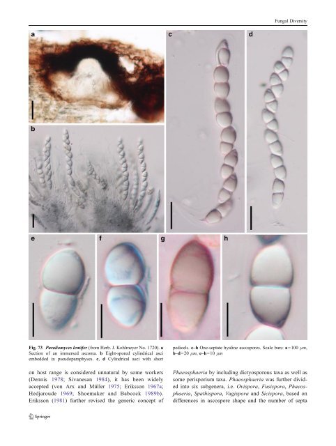
Fungal Diversity Fig. 73 Paraliomyces lentifer (from Herb. J. Kohlmeyer No. 1720). a Section of an immersed ascoma. b Eight-spored cylindrical asci embedded in pseudoparaphyses. c, d Cylindrical asci with short on host range is considered unnatural by some workers (Dennis 1978; Sivanesan 1984), it has been widely accepted (von Arx and Müller 1975; Eriksson 1967a; Hedjaroude 1969; Shoemaker and Babcock 1989b). Eriksson (1981) further revised the generic concept of pedicels. e–h One-septate hyaline ascospores. Scale bars: a=100 μm, b–d=20 μm, e–h=10 μm Phaeosphaeria by including dictyosporous taxa as well as some perisporium taxa. Phaeosphaeria was further divided into six subgenera, i.e. Ovispora, Fusispora, Phaeosphaeria, Spathispora, Vagispora and Sicispora, based on differences in ascospore shape and the number of septa
Fungal Diversity (Shoemaker and Babcock 1989b). Phaeosphaeria species are usually associated or parasitic on annual monocots, such as Cyperaceae, Juncaceae or Poaceae but have also been recorded as saprobes and on dicotyledons (e.g. P. viridella and P. vagans). Phylogenetic study The separation of Phaeosphaeria from Leptosphaeria sensu stricto was supported by phylogenetic studies based on ITS sequences. The peridium structure, pseudoparenchymatous cells in Phaeosphaeria versus scleroplectenchymatous cells in Leptosphaeria had phylogenetic significance in the distinction between these two genera, while the subgenus division was not supported by the phylogenetic results (Câmara et al. 2002; Morales et al. 1995). The familial status of both Phaeosphaeriaceae and Leptosphaeriaceae was verified by multigene phylogenetic analysis (Schoch et al. 2009; Zhang et al. 2009a). Concluding remarks Phaeosphaeria was originally thought to be a synonym of Leptosphaeria (Müller 1950; Munk1957), however, molecular analysis has shown these two genera differ with Phaeosphaeria having pseudoparenchymatous peridium, Stagonospora-like anamorph and mostly monocotyledonous hosts and Leptosphaeria having scleroplectenchymatous peridium, Phoma-like anamorph and mostly dicotyledonous hosts (Câmara et al. 2002;Schochetal.2009; Shoemaker and Babcock 1989b; Zhang et al. 2009a). It is now recognized that Phaeosphaeria is the type genus of Phaeosphaeriaceae and related genera include Entodesmium and Setomelanomma and probably Ophiosphaerella (Schoch et al. 2009; Zhangetal.2009a). Paraphaeosphaeria was introduced as an off-shoot of Phaeosphaeria and differs in ascospore shape and septation as well as anamorphic stages (Eriksson 1967a, b). Similarly, Nodulosphaeria was recently reinstated and differs from Phaeosphaeria because of setae over the apex as well as its ascospores with swelling supramedian cells and terminal appendages (Holm 1957, 1961). While the newly reinstated Phaeosphaeria was confined to monocotyledons and particularly grasses, there are now many species that have been described from dicotyledons (Farr et al. 1989). Whether these taxa form a monophyletic group needs to be investigated with fresh collections and molecular data. Phaeosphaeriopsis M.P.S. Câmara, M.E. Palm & A.W. Ramaley, Mycol. Res. 107: 519 (2003). (Phaeosphaeriaceae) Generic description Habitat terrestrial, saprobic or hemibiotrophic? Ascomata small, scattered or in small groups, immersed, globose, subglobose. Peridium thin, comprising one cell type of textura angularis. Hamathecium of dense, wide cellular pseudoparaphyses. Asci 8-spored, bitunicate, cylindrical to broadly fusoid, with a short pedicel and a small ocular chamber. Ascospores obliquely uniseriate and partially overlapping to biseriate even triseriate, cylindrical, pale brown, multi-septate, primary septum submedian, with or without constriction, verrucose or baculate. Anamorphs reported for genus: Coniothyrium-like, Phaeostagonospora (Câmara et al. 2003). Literature: Câmara et al. 2003. Type species Phaeosphaeriopsis glaucopunctata (Grev.) M.P.S. Câmara, M.E. Palm & A.W. Ramaley, Mycol. Res. 107: 519 (2003). (Fig. 75) ≡ Cryptosphaeria glaucopunctata Grev. Fl. Edin.: 362 (1824). Ascomata 120–150 μm high×140–200 μm diam., scattered, or in small groups, immersed, globose, subglobose (Fig. 75a). Peridium 10–25 μm wide, comprising one type of cells, composed of thick-walled cells of textura angularis, cells 4–9 μm diam., cell wall 2–3 μm thick, almost equal in thickness. Hamathecium of dense, wide cellular pseudoparaphyses, 3–5 μm broad. Asci (50-)60– 110×10–15 μm (x ¼ 82:3 12mm, n =10), 8-spored, bitunicate, fissitunicate dehiscence not observe, cylindrical to broadly fusoid, with a short pedicel, with a small ocular chamber (to 0.8 μm wide×1 μm high) (Fig. 75b). Ascospores 18–28×5–7.5 μm (x ¼ 23:5 6:2mm, n=10), obliquely uniseriate and partially overlapping to biseriate even triseriate, cylindrical, pale brown, 4(−5)- septate, without constriction or slightly constricted at the basal septum, the forth cell from the apex usually slightly inflated, the basal cell often longer, baculate (Fig. 75c, d, e and f). Anamorph: none reported. Material examined: UK, Epping, Sept. 1863 (E, M.C. Cooke 166, barcode: E00074286). Notes Morphology Phaeosphaeriopsis was introduced to accommodate some species of Paraphaeosphaeria based on both morphological characters and results of SSU rDNA sequence analyses (Câmara et al. 2003). Most of the Phaeosphaeriopsis species occur on the Agavaceae, although P. glaucopunctata occurs on Liliaceae (Ruscus). Phaeosphaeriopsis is characterized by having uni- or multioculate stromata and 4- or 5-septate ascospores. Although the
- Page 1 and 2:
Fungal Diversity DOI 10.1007/s13225
- Page 3 and 4:
Fungal Diversity Table 1 Major circ
- Page 5 and 6:
Fungal Diversity
- Page 7 and 8:
Fungal Diversity biocontrol agent o
- Page 9 and 10:
Fungal Diversity substrates and man
- Page 11 and 12:
Fungal Diversity 2. To investigate
- Page 13 and 14:
Fungal Diversity Table 3 (continued
- Page 15 and 16:
Fungal Diversity Table 3 (continued
- Page 17 and 18:
Fungal Diversity Table 3 (continued
- Page 19 and 20:
Fungal Diversity
- Page 21 and 22:
Fungal Diversity Fig. 2 Aigialus gr
- Page 23 and 24:
Fungal Diversity Fig. 3 Amniculicol
- Page 25 and 26:
Fungal Diversity Literature: Berkel
- Page 27 and 28:
Fungal Diversity Ascorhombispora L.
- Page 29 and 30:
Fungal Diversity
- Page 31 and 32:
Fungal Diversity Fig. 8 Astrosphaer
- Page 33 and 34:
Fungal Diversity Fig. 9 Asymmetrico
- Page 35 and 36:
Fungal Diversity Notes Morphology B
- Page 37 and 38:
Fungal Diversity Generic descriptio
- Page 39 and 40:
Fungal Diversity Anamorph: none rep
- Page 41 and 42:
Fungal Diversity Fig. 14 Bimuria no
- Page 43 and 44:
Fungal Diversity Fig. 15 Bricookea
- Page 45 and 46:
Fungal Diversity Fig. 16 Byssolophi
- Page 47 and 48:
Fungal Diversity Notes Morphology B
- Page 49 and 50:
Fungal Diversity the reaction of pe
- Page 51 and 52:
Fungal Diversity
- Page 53 and 54:
Fungal Diversity Fig. 21 Chaetomast
- Page 55 and 56:
Fungal Diversity
- Page 57 and 58:
Fungal Diversity Fig. 23 Cilioplea
- Page 59 and 60:
Fungal Diversity with one or two ve
- Page 61 and 62:
Fungal Diversity Moreau 1953; Munk
- Page 63 and 64:
Fungal Diversity Material examined:
- Page 65 and 66:
Fungal Diversity Fig. 28 Dothidotth
- Page 67 and 68:
Fungal Diversity Fig. 29 Dubitatio
- Page 69 and 70:
Fungal Diversity assigned Entodesmi
- Page 71 and 72:
Fungal Diversity fusoid to somewhat
- Page 73 and 74:
Fungal Diversity Fig. 33 Hadrospora
- Page 75 and 76:
Fungal Diversity Fig. 34 Halotthia
- Page 77 and 78:
Fungal Diversity Notes Morphology H
- Page 79 and 80:
Fungal Diversity some effused Hypox
- Page 81 and 82:
Fungal Diversity Fig. 38 Isthmospor
- Page 83 and 84:
Fungal Diversity Fig. 39 Kalmusia e
- Page 85 and 86:
Fungal Diversity ascospores were br
- Page 87 and 88:
Fungal Diversity furcate pedicel an
- Page 89 and 90: Fungal Diversity Anamorph: none rep
- Page 91 and 92: Fungal Diversity
- Page 93 and 94: Fungal Diversity Material examined:
- Page 95 and 96: Fungal Diversity Fig. 46 Lewia scro
- Page 97 and 98: Fungal Diversity Fig. 47 Lichenopyr
- Page 99 and 100: Fungal Diversity Loculohypoxylon M.
- Page 101 and 102: Fungal Diversity cells small heavil
- Page 103 and 104: Fungal Diversity upper place, septa
- Page 105 and 106: Fungal Diversity
- Page 107 and 108: Fungal Diversity (CBS 627.86) was i
- Page 109 and 110: Fungal Diversity Fig. 54 Mamillisph
- Page 111 and 112: Fungal Diversity Fig. 55 Massarina
- Page 113 and 114: Fungal Diversity phaeria as a synon
- Page 115 and 116: Fungal Diversity 5-8 μm diam., ind
- Page 117 and 118: Fungal Diversity cell wall
- Page 119 and 120: Fungal Diversity Fig. 60 Mixtura sa
- Page 121 and 122: Fungal Diversity Fig. 61 Montagnula
- Page 123 and 124: Fungal Diversity spored, bitunicate
- Page 125 and 126: Fungal Diversity Fig. 64 Murispora
- Page 127 and 128: Fungal Diversity Type species Neoph
- Page 129 and 130: Fungal Diversity brown, 8-septate,
- Page 131 and 132: Fungal Diversity Fig. 68 Ohleria mo
- Page 133 and 134: Fungal Diversity Fig. 69 Ohleriella
- Page 135 and 136: Fungal Diversity Fig. 70 Ophiobolus
- Page 137 and 138: Fungal Diversity Type species Ostro
- Page 139: Fungal Diversity
- Page 143 and 144: Fungal Diversity ium thin, composed
- Page 145 and 146: Fungal Diversity Fig. 76 Platysporo
- Page 147 and 148: Fungal Diversity Fig. 77 1 Pleomass
- Page 149 and 150: Fungal Diversity Fig. 78 Pleophragm
- Page 151 and 152: Fungal Diversity papillate, ostiola
- Page 153 and 154: Fungal Diversity Williams 1963; Mal
- Page 155 and 156: Fungal Diversity Generic descriptio
- Page 157 and 158: Fungal Diversity composed of one ce
- Page 159 and 160: Fungal Diversity Fig. 84 Saccharico
- Page 161 and 162: Fungal Diversity and nearly black a
- Page 163 and 164: Fungal Diversity dense, long trabec
- Page 165 and 166: Fungal Diversity
- Page 167 and 168: Fungal Diversity
- Page 169 and 170: Fungal Diversity Anamorphs reported
- Page 171 and 172: Fungal Diversity
- Page 173 and 174: Fungal Diversity
- Page 175 and 176: Fungal Diversity Fig. 94 Westerdyke
- Page 177 and 178: Fungal Diversity Fig. 95 Wettsteini
- Page 179 and 180: Fungal Diversity Fig. 96 Wilmia bra
- Page 181 and 182: Fungal Diversity Current name: Astr
- Page 183 and 184: Fungal Diversity spores are actuall
- Page 185 and 186: Fungal Diversity Fig. 100 Sporormie
- Page 187 and 188: Fungal Diversity
- Page 189 and 190: Fungal Diversity Fig. 102 Kriegerie
- Page 191 and 192:
Fungal Diversity Phylogenetic study
- Page 193 and 194:
Fungal Diversity Fig. 104 Zeuctomor
- Page 195 and 196:
Fungal Diversity Fig. 105 Muroia ni
- Page 197 and 198:
Fungal Diversity pseudoparenchymato
- Page 199 and 200:
Fungal Diversity Eremodothis Arx, K
- Page 201 and 202:
Fungal Diversity Type species: Macr
- Page 203 and 204:
Fungal Diversity ascospores of Plat
- Page 205 and 206:
Fungal Diversity monoceras Alcorn n
- Page 207 and 208:
Fungal Diversity tomataceae, Melano
- Page 209 and 210:
Fungal Diversity Table 4 (continued
- Page 211 and 212:
Fungal Diversity 1987b). Based on a
- Page 213 and 214:
Fungal Diversity only do so under v
- Page 215 and 216:
Fungal Diversity Dennis RWG (1968)
- Page 217 and 218:
Fungal Diversity Kirk PM, Cannon PF
- Page 219 and 220:
Fungal Diversity Saccardo PA (1880)
- Page 221:
Fungal Diversity Winter G (1887) As