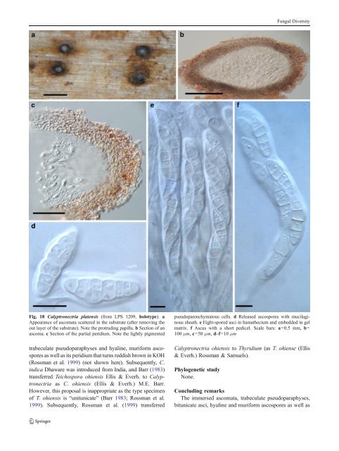
Fungal Diversity Fig. 18 Calyptronectria platensis (from LPS 1209, holotype). a Appearance of ascomata scattered in the substrate (after removing the out layer of the substrate). Note the protruding papilla. b Section of an ascoma. c Section of the partial peridium. Note the lightly pigmented trabeculate pseudoparaphyses and hyaline, muriform ascospores as well as its peridium that turns reddish brown in KOH (Rossman et al. 1999) (not shown here). Subsequently, C. indica Dhaware was introduced from India, and Barr (1983) transferred Teichospora ohiensis Ellis & Everh. to Calyptronectria as C. ohiensis (Ellis & Everh.) M.E. Barr. However, this proposal is inappropriate as the type specimen of T. ohiensis is “unitunicate” (Barr 1983; Rossman et al. 1999). Subsequently, Rossman et al. (1999) transferred pseudoparenchymatous cells. d Released ascospores with mucilaginous sheath. e Eight-spored asci in hamathecium and embedded in gel matrix. f Ascus with a short pedicel. Scale bars: a=0.5 mm, b= 100 μm, c=50 μm, d–f=10 μm Calyptronectria ohiensis to Thyridium (as T. ohiense (Ellis & Everh.) Rossman & Samuels). Phylogenetic study None. Concluding remarks The immersed ascomata, trabeculate pseudoparaphyses, bitunicate asci, hyaline and muriform ascospores as well as
Fungal Diversity the reaction of peridium to KOH (turns reddish brown) make it distinguishable from all other reported genera (Rossman et al. 1999). Thus Calyptronectria is a morphologically well defined genus. Carinispora K.D. Hyde, J. Linn. Soc., Bot. 110: 97 (1992). (<strong>Pleosporales</strong>, genera incertae sedis) Generic description Habitat marine, saprobic. One or two ascomata per stroma. Ascomata scattered or in small groups, developing beneath the host epidermis, erumpent, lenticular, ostiolate, lacking periphyses. Peridium pale brown, composed of thin-walled elongated cells at the sides and thick-walled cells of textura epidermoidea at the base. Hamathecium of dense, long filliform pseudoparaphyses, embedded in mucilage, anastomosing between and above the asci, rarely septate. Asci 8-spored, bitunicate, fissitunicate, clavate to cylindrical, with a short furcate pedicel, apex with an ocular chamber and apical ring. Ascospores biseriate, narrowly fusoid, yellow to pale brown, multi-septate, constricted at the septa, the two central cells being the largest, surrounded by a gelatinous sheath. Anamorphs reported for genus: none. Literature: Hyde 1992a, 1994b. Type species Carinispora nypae K.D. Hyde, J. Linn. Soc., Bot. 110: 99 (1992). (Fig. 19) One or two ascomata per stroma. Ascomata up to 0.8 mm diam., scattered or in small groups, developing beneath the host epidermis, crust-like, as circular spots, wall brown, with a small central ostiole, in section 225–285 μm high×510– 750 μm diam., lenticular, ostiolar canal lacking periphyses (Fig. 19a and b). Peridium 35–45 μm wide at sides, pale brown, at sides composed of a thin layer of thin-walled elongate cells, fusing with the stromatic tissue and host cells, at the base composed of thick-walled cells, forming a textura epidermoidea and fusing with host cells. A wedge of pale brown hyphae forming a textura porrecta is present at the rim (Fig. 19c). Hamathecium of dense, long filliform pseudoparaphyses 1–3 μm broad, embedded in mucilage, anastomosing between and above the asci, rarely septate. Asci 142–207×14.2–19.8 μm, 8-spored, bitunicate, fissitunicate, clavate to cylindrical, with a furcate pedicel, up to 40 μm long, apex with an ocular chamber and apical ring (to 2 μm wide×3 μm high, J-), developing from ascogenous tissue at the base of the ascocarp (Fig. 19d, e, f, g and h). Ascospores 42–66×7–10.6 μm, biseriate, narrowly fusoid with broadly to narrowly rounded ends, somewhat curved, yellow to pale brown, yellow in mass, 7-8-septate, constricted at the septa, the two central cells being the largest, surrounded by a gelatinous sheath; the sheath has a central “spine” and curved polar extrusions (Fig. 19i and j). Anamorph: none reported. Material examined: BRUNEI DARUSSALAM, Tungit Api Api mangrove, from decaying intertidal fronds of Nypa fruticans Wurmb., 14 Apr. 1987, K.D. Hyde (BRIP 17106, holotype). Notes Morphology Carinispora is distinguished from Phaeosphaeria by its saprobic life style and lenticular ascomata formed under the host epidermis, peridium structure and sheath surrounding the ascospores (Hyde 1992a, 1994b). Two species were reported, i.e. C. nypae and C. velatispora K.D. Hyde. Phylogenetic study Suetrong et al. (2009) could not resolve Carinispora nypae in a phylogeny based on four genes. Concluding remarks Both Carinispora nypae and C. velatispora are reported as marine fungi, which should be taken into consideration for their familial placement. Caryosporella Kohlm., Proc. Indian Acad. Sci., Pl. Sci. 94: 355 (1985). (?Melanommataceae) Generic description Habitat marine, saprobic. Ascomata densely scattered or gregarious, superficial, subglobose, black, papillate, ostiolate, periphysate, carbonaceous. Peridium carbonaceous. Hamathecium of dense, long trabeculate pseudoparaphyses, anastomosing and branching above the asci. Asci 8-spored, bitunicate, fissitunicate, cylindrical. Ascospores ellipsoidal to broadly fusoid with narrowly hyaline rounded ends, deep reddish brown, thickwalled, 1-septate with hyaline germ pore at each end. Anamorphs reported for genus: suspected spermatia (Kohlmeyer 1985). Literature: Eriksson 2006; Kohlmeyer 1985; Lumbsch and Huhndorf 2007. Type species Caryosporella rhizophorae Kohlm., Proc. Indian Acad. Sci., Pl. Sci. 94: 356 (1985). (Fig. 20) Ascomata 0.8–1.1 mm high×0.9–1.2 mm diam., densely scattered or gregarious, superficial with a flattened base, not easily removed from the host surface, subglobose, black, short
- Page 1 and 2: Fungal Diversity DOI 10.1007/s13225
- Page 3 and 4: Fungal Diversity Table 1 Major circ
- Page 5 and 6: Fungal Diversity
- Page 7 and 8: Fungal Diversity biocontrol agent o
- Page 9 and 10: Fungal Diversity substrates and man
- Page 11 and 12: Fungal Diversity 2. To investigate
- Page 13 and 14: Fungal Diversity Table 3 (continued
- Page 15 and 16: Fungal Diversity Table 3 (continued
- Page 17 and 18: Fungal Diversity Table 3 (continued
- Page 19 and 20: Fungal Diversity
- Page 21 and 22: Fungal Diversity Fig. 2 Aigialus gr
- Page 23 and 24: Fungal Diversity Fig. 3 Amniculicol
- Page 25 and 26: Fungal Diversity Literature: Berkel
- Page 27 and 28: Fungal Diversity Ascorhombispora L.
- Page 29 and 30: Fungal Diversity
- Page 31 and 32: Fungal Diversity Fig. 8 Astrosphaer
- Page 33 and 34: Fungal Diversity Fig. 9 Asymmetrico
- Page 35 and 36: Fungal Diversity Notes Morphology B
- Page 37 and 38: Fungal Diversity Generic descriptio
- Page 39 and 40: Fungal Diversity Anamorph: none rep
- Page 41 and 42: Fungal Diversity Fig. 14 Bimuria no
- Page 43 and 44: Fungal Diversity Fig. 15 Bricookea
- Page 45 and 46: Fungal Diversity Fig. 16 Byssolophi
- Page 47: Fungal Diversity Notes Morphology B
- Page 51 and 52: Fungal Diversity
- Page 53 and 54: Fungal Diversity Fig. 21 Chaetomast
- Page 55 and 56: Fungal Diversity
- Page 57 and 58: Fungal Diversity Fig. 23 Cilioplea
- Page 59 and 60: Fungal Diversity with one or two ve
- Page 61 and 62: Fungal Diversity Moreau 1953; Munk
- Page 63 and 64: Fungal Diversity Material examined:
- Page 65 and 66: Fungal Diversity Fig. 28 Dothidotth
- Page 67 and 68: Fungal Diversity Fig. 29 Dubitatio
- Page 69 and 70: Fungal Diversity assigned Entodesmi
- Page 71 and 72: Fungal Diversity fusoid to somewhat
- Page 73 and 74: Fungal Diversity Fig. 33 Hadrospora
- Page 75 and 76: Fungal Diversity Fig. 34 Halotthia
- Page 77 and 78: Fungal Diversity Notes Morphology H
- Page 79 and 80: Fungal Diversity some effused Hypox
- Page 81 and 82: Fungal Diversity Fig. 38 Isthmospor
- Page 83 and 84: Fungal Diversity Fig. 39 Kalmusia e
- Page 85 and 86: Fungal Diversity ascospores were br
- Page 87 and 88: Fungal Diversity furcate pedicel an
- Page 89 and 90: Fungal Diversity Anamorph: none rep
- Page 91 and 92: Fungal Diversity
- Page 93 and 94: Fungal Diversity Material examined:
- Page 95 and 96: Fungal Diversity Fig. 46 Lewia scro
- Page 97 and 98: Fungal Diversity Fig. 47 Lichenopyr
- Page 99 and 100:
Fungal Diversity Loculohypoxylon M.
- Page 101 and 102:
Fungal Diversity cells small heavil
- Page 103 and 104:
Fungal Diversity upper place, septa
- Page 105 and 106:
Fungal Diversity
- Page 107 and 108:
Fungal Diversity (CBS 627.86) was i
- Page 109 and 110:
Fungal Diversity Fig. 54 Mamillisph
- Page 111 and 112:
Fungal Diversity Fig. 55 Massarina
- Page 113 and 114:
Fungal Diversity phaeria as a synon
- Page 115 and 116:
Fungal Diversity 5-8 μm diam., ind
- Page 117 and 118:
Fungal Diversity cell wall
- Page 119 and 120:
Fungal Diversity Fig. 60 Mixtura sa
- Page 121 and 122:
Fungal Diversity Fig. 61 Montagnula
- Page 123 and 124:
Fungal Diversity spored, bitunicate
- Page 125 and 126:
Fungal Diversity Fig. 64 Murispora
- Page 127 and 128:
Fungal Diversity Type species Neoph
- Page 129 and 130:
Fungal Diversity brown, 8-septate,
- Page 131 and 132:
Fungal Diversity Fig. 68 Ohleria mo
- Page 133 and 134:
Fungal Diversity Fig. 69 Ohleriella
- Page 135 and 136:
Fungal Diversity Fig. 70 Ophiobolus
- Page 137 and 138:
Fungal Diversity Type species Ostro
- Page 139 and 140:
Fungal Diversity
- Page 141 and 142:
Fungal Diversity (Shoemaker and Bab
- Page 143 and 144:
Fungal Diversity ium thin, composed
- Page 145 and 146:
Fungal Diversity Fig. 76 Platysporo
- Page 147 and 148:
Fungal Diversity Fig. 77 1 Pleomass
- Page 149 and 150:
Fungal Diversity Fig. 78 Pleophragm
- Page 151 and 152:
Fungal Diversity papillate, ostiola
- Page 153 and 154:
Fungal Diversity Williams 1963; Mal
- Page 155 and 156:
Fungal Diversity Generic descriptio
- Page 157 and 158:
Fungal Diversity composed of one ce
- Page 159 and 160:
Fungal Diversity Fig. 84 Saccharico
- Page 161 and 162:
Fungal Diversity and nearly black a
- Page 163 and 164:
Fungal Diversity dense, long trabec
- Page 165 and 166:
Fungal Diversity
- Page 167 and 168:
Fungal Diversity
- Page 169 and 170:
Fungal Diversity Anamorphs reported
- Page 171 and 172:
Fungal Diversity
- Page 173 and 174:
Fungal Diversity
- Page 175 and 176:
Fungal Diversity Fig. 94 Westerdyke
- Page 177 and 178:
Fungal Diversity Fig. 95 Wettsteini
- Page 179 and 180:
Fungal Diversity Fig. 96 Wilmia bra
- Page 181 and 182:
Fungal Diversity Current name: Astr
- Page 183 and 184:
Fungal Diversity spores are actuall
- Page 185 and 186:
Fungal Diversity Fig. 100 Sporormie
- Page 187 and 188:
Fungal Diversity
- Page 189 and 190:
Fungal Diversity Fig. 102 Kriegerie
- Page 191 and 192:
Fungal Diversity Phylogenetic study
- Page 193 and 194:
Fungal Diversity Fig. 104 Zeuctomor
- Page 195 and 196:
Fungal Diversity Fig. 105 Muroia ni
- Page 197 and 198:
Fungal Diversity pseudoparenchymato
- Page 199 and 200:
Fungal Diversity Eremodothis Arx, K
- Page 201 and 202:
Fungal Diversity Type species: Macr
- Page 203 and 204:
Fungal Diversity ascospores of Plat
- Page 205 and 206:
Fungal Diversity monoceras Alcorn n
- Page 207 and 208:
Fungal Diversity tomataceae, Melano
- Page 209 and 210:
Fungal Diversity Table 4 (continued
- Page 211 and 212:
Fungal Diversity 1987b). Based on a
- Page 213 and 214:
Fungal Diversity only do so under v
- Page 215 and 216:
Fungal Diversity Dennis RWG (1968)
- Page 217 and 218:
Fungal Diversity Kirk PM, Cannon PF
- Page 219 and 220:
Fungal Diversity Saccardo PA (1880)
- Page 221:
Fungal Diversity Winter G (1887) As