

Pleosporales - CBS - KNAW
Pleosporales - CBS - KNAW
Pleosporales - CBS - KNAW

You also want an ePaper? Increase the reach of your titles
YUMPU automatically turns print PDFs into web optimized ePapers that Google loves.
Fungal Diversity<br />
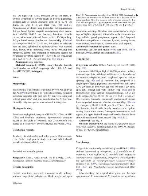
190 μm high (Fig. 101a). Peridium 20–33 μm thick, 1-<br />
layered, composed of several layers of heavily pigmented,<br />
elongate cells of textura angularis, cells up to 6.3×5 μm<br />
diam., cell wall 1–1.5 μm thick (Fig. 101b and c).<br />
Hamathecium of dense, long trabeculate pseudoparaphyses<br />
1–2 μm broad, hyaline, aseptate, decomposing when mature.<br />
Asci 165–220×33–42.5 μm, 8-spored, bitunicate, broadly<br />
clavate, with a small, thin and furcate pedicel, 35–50 μm long,<br />
3–5 μm broad, ocular chamber not observed (Fig. 101d and<br />
e). Ascospores 68–93×12.5–16 μm, 3–4 seriate to uniseriate<br />
near the base, cylindrical to cylindro-clavate with rounded<br />
ends, brown, (6-)7 transverse septa, easily breaking into<br />
partspores, central cells triangular in transverse section but<br />
rectangular in vertical section, 14–16.5×8–10 μm long, apical<br />
cells 12.5–15×11.5–17.5 μm long (Fig. 101f and g).<br />
Anamorph: none reported.<br />
Material examined: SPAIN, Canary Islands, Tenerifa<br />
Las Canadas, on rabbit? droppings, Mar. 1986, J.A. von<br />
Arx (H<strong>CBS</strong> 9812, holotype).<br />
Notes<br />
Morphology<br />
Spororminula was formally established by von Arx and van<br />
der Aa (1987) according to its “ostiolate ascomata, elongated<br />
ascospore separated into part cells by transverse septa and<br />
without germ slits”, and was monotypified by S. tenerifae.<br />
Currently, only one species was included in this genus.<br />
Phylogenetic study<br />
Based on a phylogenetic analysis of ITS-nLSU rDNA, mtSSU<br />
rDNA and ß-tubulin sequences, Spororminula tenerifae<br />
nested in the clade of Preussia, thus Spororminula was<br />
treated as a synonym of Preussia (Kruys and Wedin 2009).<br />
Concluding remarks<br />
To clarify its relationship with other genera of Sporormiaceae,<br />
further phylogenetic study is needed, which should<br />
include additional related taxa.<br />
Excluded and doubtful genera<br />
Kriegeriella Höhn., Annls mycol. 16: 39 (1918). (Dothideomycetes,<br />
families incertae sedis, Microthyriaceae)<br />
Generic description<br />
Habitat terrestrial, saprobic? Ascomata small, solitary,<br />
scattered, superficial, subglobose, black, roughened, apex<br />
Fig. 101 Spororminula tenerifae (from H<strong>CBS</strong> 9812, holotype). a b<br />
Appearance of ascomata on the host surface. b, c Sections of the<br />
partial peridium. Note the elongate cells of textura angularis. d, e<br />
Asci with thin pedicels. f, g Ascospores, which may break into part<br />
spores. Scale bars: a=0.5 mm, b=100 μm, c=50 μm, d–g=20 μm<br />
no obvious opening. Peridium thin, composed of a single<br />
type of lightly pigmented thin-walled cells. Hamathecium<br />
long cellular pseudoparaphyses, septate. Asci 8-spored,<br />
bitunicate, obpyriform. Ascospores hyaline, turning brown<br />
when mature, multi-septate, constricted at each septum.<br />
Anamorphs reported for genus: none.<br />
Literature: von Arx and Müller 1975; Barr1975, 1987b;<br />
Eriksson 2006; Lumbsch and Huhndorf 2007.<br />
Type species<br />
Kriegeriella mirabilis Höhn., Annls mycol. 16: 39 (1918)<br />
(Fig. 102)<br />
Ascomata 100–120 μm high×150–220 μm diam., solitary,<br />
scattered, superficial, with basal wall flattened on the surface of<br />
the substrate, subglobose, black, roughened, apex no obvious<br />
opening (Fig. 102a and e). Peridium thin, composed of a<br />
single type of lightly pigmented thin-walled cells, cells up to<br />
12×5 μm diam. in front view, cell wall less than 1 μm thick,<br />
apex cells smaller and walls thicker (Fig. 102a and f).<br />
Hamathecium long cellular pseudoparaphyses, 1.5–2 μm<br />
wide, septate. Asci 65–85×31–36 μm (x ¼ 63:1 33mm, n=<br />
10), 8-spored, bitunicate, fissitunicate undetermined, obpyriform,<br />
no pedicel, no ocular chamber was seen (Fig. 102c and<br />
g). Ascospores 28–37.5×8–11 μm (x ¼ 32:6 10mm, n=<br />
10), 8-seriate, fusoid with broadly rounded ends, hyaline,<br />
turning brown when mature, 5–6-septate, constricted at each<br />
septum, the upper first-cell longer and broader than the lower<br />
ones with semi-round shape, smooth (Fig. 102d, h, i).<br />
Anamorph: see Fig. b.<br />
Material examined: On the leaves of Faulenden nadeln<br />
von Pinus silvestris, bei Roñigstein, Sept. 1896, W. Rueges.<br />
(S reg. nr F12638, isolectotype).<br />
Notes<br />
Morphology<br />
Kriegeriella was formally established by von Höhnel (1918b)<br />
and was represented by two species, i.e. K. mirabilis and K.<br />
transiens; it was typified by K. mirabilis and assigned to<br />
Microthyriaceae. Subsequently, Kriegeriella was assigned to<br />
the subfamily of Aulographiodeae (Microthyriaceae)<br />
(Batista et al. 1959), Asterinaceae (Hemisphaeriales) (Luttrell<br />
1973) and Pseudosphaeriaceae (Dothideales) (Barr<br />
1975).<br />
After checking the original description and the type<br />
specimens of K. mirabilis and K. transiens, no significant


