

Pleosporales - CBS - KNAW
Pleosporales - CBS - KNAW
Pleosporales - CBS - KNAW

Create successful ePaper yourself
Turn your PDF publications into a flip-book with our unique Google optimized e-Paper software.
Fungal Diversity<br />
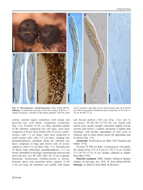
Fig. 17 Byssosphaeria schiedermayriana (from K(M):108784,<br />
holotype). a Superficial ascomata on the host surface. b Brown, 1-<br />
septate ascospores. c Section of the lateral peridium. Note the outer<br />
textura angularis and inner textura epidermoidea cells. d, e Furcate<br />
asci with a long pedicel. f Dehiscent ascus. Scale bars: a=0.5 mm, c=<br />
50 μm, b, d–f=15 μm<br />
ostiole, ostiolar region sometimes with orange and<br />
greenish tint, wall black, roughened, coriaceous<br />
(Fig. 17a). Peridium 55–85 μm thick, peridium outside<br />
of the substrate comprising two cell types, outer layer<br />
composed of brown thick-walled cells of textura epidermoidea,<br />
cells 1–3 μm diam., inner layer composed of<br />
small hyaline cells, cells 3–5 μm diam., merging into<br />
pseudoparaphyses; peridium inside the substrate one<br />
layer, composed of large pale brown cells of textura<br />
angularis, cells6–13 μm diam. (Fig. 17c). Hamathecium<br />
of dense, long trabeculate pseudoparaphyses, 1–2 μm<br />
broad, embedded in mucilage, anastomosing between and<br />
above the asci. Asci 90–120(−148)×10–14 μm, 8-spored,<br />
bitunicate, fissitunicate, cylindro-clavate to clavate,<br />
biseriate above and uniseriate below, pedicel 15–20<br />
(−53) μm long, the immature asci usually with longer<br />
and furcate pedicel (−68 μm) (Fig. 17d,e and f).<br />
Ascospores 29–34(−38)×5.5–8(−10) μm, fusoid with<br />
narrow ends, mostly straight, sometimes slightly curved,<br />
smooth, pale brown, 1-septate, becoming 3-septate after<br />
discharge, with hyaline appendages at each acute to<br />
subacute end; in some mature spores the appendage may<br />
be absent (Fig. 17b).<br />
Anamorph: Pyrenochaeta sp. (Barr 1984; Samuels and<br />
Müller 1978).<br />
Pycnidia 70–500 μm diam. Conidiogenous cells phialidic,<br />
lining cavity, 5–8×4–6 μm to 5–10×3–6 μm. Conidia<br />
2.5–3.5(−4)×1.5–2(−3) μm, hyaline, ellipsoid or subglobose<br />
(Barr 1984).<br />
Material examined: ERIE, Dublin, Glasnevin Botanic<br />
Garden, on old rope, Jun. 1872, W. Keit (K(M):108784,<br />
holotype, asSphaeria keitii Berk. & Broome).


