Fungal Diversity transverse septa, 1 longitudinal septum in each central cell, 1 oblique septum in each end cell, constricted at all septa, granulate, with a sheath 2–3 μm wide (as reported in Shoemaker and Babcock 1992) (Fig.76f, g and h). Anamorph: none reported. Material examined: GERMANY, Budenheim, Leopold Fuckel, Nassau’s Flora, on old paper (G NASSAU: 210558 (a), as Sphaeria chartarum Wallr., type). Notes Morphology Platysporoides was introduced as a subgenus of Pleospora by Wehmeyer (1961) and was typified by Pleospora chartarum. Shoemaker and Babcock (1992) raised Platysporoides to generic rank and placed it in the Pleosporaceae based on its “applanodictyospore” and “terete pored beak of the ascomata”. Currently, eleven species are included in this genus (Shoemaker and Babcock 1992). Another comparable pleosporalean family is Diademaceae, which is distinguished from Platysporoides by its ascoma opening as “an intraepidermal discoid lid” (Shoemaker and Babcock 1992). Phylogenetic study None. Concluding remarks Aigialus grandis is another pleosporalean fungus with flattened and muriform ascospores as well as papilla and ostioles, which belongs to Aigialaceae, a phylogenetically well supported marine family (Suetrong et al. 2009). Thus, it is highly likely that flattened and muriform ascospores are of little phylogenetic significance. Pleomassaria Speg., Anal. Soc. cient. argent. 9: 192 (1880). (Pleomassariaceae) Generic description Habitat terrestrial, saprobic. Ascomata medium to large, solitary, scattered, or in small groups, immersed, erumpent by a minute slit or a small conical swelling in the bark, flattened, papillate, ostiolate. Hamathecium of dense, cellular pseudoparaphyses, embedded in mucilage. Asci bitunicate, fissitunicate, broadly cylindrical to broadly cylindro-clavate, with a short, thick pedicel. Ascospores muriform, brown, constricted at the septa. Anamorphs reported for genus: Prosthemium and Shearia (Barr 1982b; Sivanesan 1984). Literature: Barr 1982b, 1990b, 1993a; Clements and Shear 1931; Eriksson 2006; Lumbsch and Huhndorf 2007; Shoemaker and LeClair 1975; Sivanesan 1984; Tanaka et al. 2005. Type species Pleomassaria siparia (Berk. & Broome) Sacc., Syll. fung. 2: 239 (1883) (Fig. 77) ≡ Sphaeria siparia Berk. & Broome, Ann. Mag. nat. Hist., Ser. 2 9: 321 (1852). Ascomata 150–410 μm high×440–740 μm diam., solitary, scattered, or in small groups, immersed, erumpent by a minute slit or a small conical swelling in the bark, depressed globose, papillalte, ostiolate (Fig. 77a). Peridium 45–60 μm wide, thicker at the apex, thinner at the base, 1-layered, composed of small pigmented thickwalled compressed cells, cells ca. 15×3μm diam., cell wall 2–3.5 μm thick, apex cells larger, base composed of small pigmented thick-walled cells of textura angularis, ca. 5μm diam. (Fig. 77b). Hamathecium of dense, cellular pseudoparaphyses, 1–2 μm broad, embedded in mucilage, anastomosing or branching not observed. Asci 180–250× 28–42 μm (x¼206:3 36:8mm, n=10), 8-spored, bitunicate, fissitunicate, broadly cylindrical to broadly cylindroclavate, with a short, thick pedicel, 15–45 μm long, with inconspicuous ocular chamber (Fig. 77c and d). Ascospores 45–58×12.5–17.5 μm (x ¼ 50:5 14:8mm, n=10), biseriate, narrowly oblong with broadly to narrowly rounded ends, brown, muriform with 5–8 transverse septa and 1–2 vertical septa in some cells, smooth to verrucose, constricted at the septa, surrounded by a mucilaginous sheath (Fig. 77e, f and g). Anamorph: Prosthemium betulinum Kunze (Sivanesan 1984). Conidia to 120 μm diam., with 3–5 arms, each arm 3–5- septate, 40–55×13–16 μm, connected to a central cell (Fig. 77h, i and j). Material examined: UK, Wiltshire, Spye Park, on branch of Betulina with Hendersonia polycystis Berk., et Br. leg. C.E. Broome, 1850? (BR, type). Notes Morphology Pleomassaria as characterized by Barr (1982b) has medium- to large-sized, immersed ascomata, cellular pseudoparaphyses, clavate to oblong asci and large, muriform ascospores (Barr 1982b; Sivanesan 1984). The muriform and somewhat asymmetrical ascospores with a submedian primary septum distinguish Pleomassaria from Asteromassaria in the family Pleomassariaceae, while in Splanchnonema ascospores have distinct bipolar asymmetry. Barr (1982b) included five North American species in the genus, while Kirk et al. (2008) listedfourspecies.
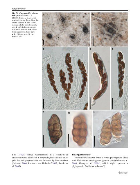
Fungal Diversity Fig. 76 Platysporoides chartarum (from G NASSAU: 210558, type). a, b Ascomata scattered among fibers. Note the central ostioles. c Asci in numerous cellular pseudoparaphyses. d, e Cylindro-clavate asci with short pedicels. f–h. Muriform ascospores. Scale bars: a, b=200 μm, c–e=20 μm, f–h=10 μm Barr (1993a) treated Pleomassaria as a synonym of Splanchnonema based on a morphological cladistic analysis, but this proposal was not followed by later workers (Eriksson 2006; Lumbsch and Huhndorf 2007; Tanaka et al. 2005). Phylogenetic study Pleomassaria siparia forms a robust phylogenetic clade with Melanomma pulvis-pyrius (generic type) (Schoch et al. 2009; Zhang et al. 2009a), which might represent a phylogenetic family (or suborder?).
- Page 1 and 2:
Fungal Diversity DOI 10.1007/s13225
- Page 3 and 4:
Fungal Diversity Table 1 Major circ
- Page 5 and 6:
Fungal Diversity
- Page 7 and 8:
Fungal Diversity biocontrol agent o
- Page 9 and 10:
Fungal Diversity substrates and man
- Page 11 and 12:
Fungal Diversity 2. To investigate
- Page 13 and 14:
Fungal Diversity Table 3 (continued
- Page 15 and 16:
Fungal Diversity Table 3 (continued
- Page 17 and 18:
Fungal Diversity Table 3 (continued
- Page 19 and 20:
Fungal Diversity
- Page 21 and 22:
Fungal Diversity Fig. 2 Aigialus gr
- Page 23 and 24:
Fungal Diversity Fig. 3 Amniculicol
- Page 25 and 26:
Fungal Diversity Literature: Berkel
- Page 27 and 28:
Fungal Diversity Ascorhombispora L.
- Page 29 and 30:
Fungal Diversity
- Page 31 and 32:
Fungal Diversity Fig. 8 Astrosphaer
- Page 33 and 34:
Fungal Diversity Fig. 9 Asymmetrico
- Page 35 and 36:
Fungal Diversity Notes Morphology B
- Page 37 and 38:
Fungal Diversity Generic descriptio
- Page 39 and 40:
Fungal Diversity Anamorph: none rep
- Page 41 and 42:
Fungal Diversity Fig. 14 Bimuria no
- Page 43 and 44:
Fungal Diversity Fig. 15 Bricookea
- Page 45 and 46:
Fungal Diversity Fig. 16 Byssolophi
- Page 47 and 48:
Fungal Diversity Notes Morphology B
- Page 49 and 50:
Fungal Diversity the reaction of pe
- Page 51 and 52:
Fungal Diversity
- Page 53 and 54:
Fungal Diversity Fig. 21 Chaetomast
- Page 55 and 56:
Fungal Diversity
- Page 57 and 58:
Fungal Diversity Fig. 23 Cilioplea
- Page 59 and 60:
Fungal Diversity with one or two ve
- Page 61 and 62:
Fungal Diversity Moreau 1953; Munk
- Page 63 and 64:
Fungal Diversity Material examined:
- Page 65 and 66:
Fungal Diversity Fig. 28 Dothidotth
- Page 67 and 68:
Fungal Diversity Fig. 29 Dubitatio
- Page 69 and 70:
Fungal Diversity assigned Entodesmi
- Page 71 and 72:
Fungal Diversity fusoid to somewhat
- Page 73 and 74:
Fungal Diversity Fig. 33 Hadrospora
- Page 75 and 76:
Fungal Diversity Fig. 34 Halotthia
- Page 77 and 78:
Fungal Diversity Notes Morphology H
- Page 79 and 80:
Fungal Diversity some effused Hypox
- Page 81 and 82:
Fungal Diversity Fig. 38 Isthmospor
- Page 83 and 84:
Fungal Diversity Fig. 39 Kalmusia e
- Page 85 and 86:
Fungal Diversity ascospores were br
- Page 87 and 88:
Fungal Diversity furcate pedicel an
- Page 89 and 90:
Fungal Diversity Anamorph: none rep
- Page 91 and 92:
Fungal Diversity
- Page 93 and 94: Fungal Diversity Material examined:
- Page 95 and 96: Fungal Diversity Fig. 46 Lewia scro
- Page 97 and 98: Fungal Diversity Fig. 47 Lichenopyr
- Page 99 and 100: Fungal Diversity Loculohypoxylon M.
- Page 101 and 102: Fungal Diversity cells small heavil
- Page 103 and 104: Fungal Diversity upper place, septa
- Page 105 and 106: Fungal Diversity
- Page 107 and 108: Fungal Diversity (CBS 627.86) was i
- Page 109 and 110: Fungal Diversity Fig. 54 Mamillisph
- Page 111 and 112: Fungal Diversity Fig. 55 Massarina
- Page 113 and 114: Fungal Diversity phaeria as a synon
- Page 115 and 116: Fungal Diversity 5-8 μm diam., ind
- Page 117 and 118: Fungal Diversity cell wall
- Page 119 and 120: Fungal Diversity Fig. 60 Mixtura sa
- Page 121 and 122: Fungal Diversity Fig. 61 Montagnula
- Page 123 and 124: Fungal Diversity spored, bitunicate
- Page 125 and 126: Fungal Diversity Fig. 64 Murispora
- Page 127 and 128: Fungal Diversity Type species Neoph
- Page 129 and 130: Fungal Diversity brown, 8-septate,
- Page 131 and 132: Fungal Diversity Fig. 68 Ohleria mo
- Page 133 and 134: Fungal Diversity Fig. 69 Ohleriella
- Page 135 and 136: Fungal Diversity Fig. 70 Ophiobolus
- Page 137 and 138: Fungal Diversity Type species Ostro
- Page 139 and 140: Fungal Diversity
- Page 141 and 142: Fungal Diversity (Shoemaker and Bab
- Page 143: Fungal Diversity ium thin, composed
- Page 147 and 148: Fungal Diversity Fig. 77 1 Pleomass
- Page 149 and 150: Fungal Diversity Fig. 78 Pleophragm
- Page 151 and 152: Fungal Diversity papillate, ostiola
- Page 153 and 154: Fungal Diversity Williams 1963; Mal
- Page 155 and 156: Fungal Diversity Generic descriptio
- Page 157 and 158: Fungal Diversity composed of one ce
- Page 159 and 160: Fungal Diversity Fig. 84 Saccharico
- Page 161 and 162: Fungal Diversity and nearly black a
- Page 163 and 164: Fungal Diversity dense, long trabec
- Page 165 and 166: Fungal Diversity
- Page 167 and 168: Fungal Diversity
- Page 169 and 170: Fungal Diversity Anamorphs reported
- Page 171 and 172: Fungal Diversity
- Page 173 and 174: Fungal Diversity
- Page 175 and 176: Fungal Diversity Fig. 94 Westerdyke
- Page 177 and 178: Fungal Diversity Fig. 95 Wettsteini
- Page 179 and 180: Fungal Diversity Fig. 96 Wilmia bra
- Page 181 and 182: Fungal Diversity Current name: Astr
- Page 183 and 184: Fungal Diversity spores are actuall
- Page 185 and 186: Fungal Diversity Fig. 100 Sporormie
- Page 187 and 188: Fungal Diversity
- Page 189 and 190: Fungal Diversity Fig. 102 Kriegerie
- Page 191 and 192: Fungal Diversity Phylogenetic study
- Page 193 and 194: Fungal Diversity Fig. 104 Zeuctomor
- Page 195 and 196:
Fungal Diversity Fig. 105 Muroia ni
- Page 197 and 198:
Fungal Diversity pseudoparenchymato
- Page 199 and 200:
Fungal Diversity Eremodothis Arx, K
- Page 201 and 202:
Fungal Diversity Type species: Macr
- Page 203 and 204:
Fungal Diversity ascospores of Plat
- Page 205 and 206:
Fungal Diversity monoceras Alcorn n
- Page 207 and 208:
Fungal Diversity tomataceae, Melano
- Page 209 and 210:
Fungal Diversity Table 4 (continued
- Page 211 and 212:
Fungal Diversity 1987b). Based on a
- Page 213 and 214:
Fungal Diversity only do so under v
- Page 215 and 216:
Fungal Diversity Dennis RWG (1968)
- Page 217 and 218:
Fungal Diversity Kirk PM, Cannon PF
- Page 219 and 220:
Fungal Diversity Saccardo PA (1880)
- Page 221:
Fungal Diversity Winter G (1887) As