

Pleosporales - CBS - KNAW
Pleosporales - CBS - KNAW
Pleosporales - CBS - KNAW

You also want an ePaper? Increase the reach of your titles
YUMPU automatically turns print PDFs into web optimized ePapers that Google loves.
Fungal Diversity<br />
hyaline small cells of textura epidermoidea, 2–4 μm diam.,<br />
cell wall 1–3 μm thick, interspersed with interwoven<br />
mycelium in places (Fig. 89b and c). Hamathecium of dense,<br />
broadly trabeculate pseudoparaphyses 1–2 μm broad, anastomosing<br />
between and above the asci (Fig. 89d). Asci 140–190<br />
(−205)×12.5–15(−17.5) μm (x ¼ 164 14:3mm, n=10), 8-<br />
spored, bitunicate, cylindrical, with a short, furcate pedicel,<br />
20–45 μm long, and an inconspicuous ocular chamber (to<br />
2 μm wide×1 μm high) (Fig. 89d and e). Ascospores 20–25×<br />
10–12 μm (x ¼ 22:1 10:3mm, n=10), obliquely uniseriate<br />
and partially overlapping, broadly ellipsoid with rounded<br />
ends, hyaline, becoming pale brown when mature, 1-septate,<br />
constricted at the median septum, smooth (Fig. 89f).<br />
Anamorph: none reported.<br />
Material examined: CHINA, Kansu Prov., between<br />
Scharakuto and Kweite, on rotten stems of Salsola gemmascens<br />
Pall., 25 Jul. 1935, G. Fenzel 2400 (W 16366, type).<br />
Notes<br />
Morphology<br />
Sinodidymella was formally established by Yue and<br />
Eriksson (1985) as they noticed that Amphididymella<br />
verrucosa Petr. was not congeneric with the generic type,<br />
A. adeana Petr., which is a pyrenolichen. Thus a new<br />
monotypic genus, Sinodidymella was introduced to accommodate<br />
it. The most outstanding morphological character of<br />
Sinodidymella is its radial ridges, which are somewhat<br />
comparable with that of Lophiostoma rugulosum Yin.<br />
Zhang, J. Fourn. & K.D. Hyde, although their pseudoparaphyses<br />
are dissimilar. Lophiostoma rugulosum has “tightly<br />
aggregated cellular pseudoparaphyses” and “apically ending<br />
into bunches of clavate cells” (Zhang et al. 2009b).<br />
Phylogenetic study<br />
None.<br />
Concluding remarks<br />
The radial ridges have little phylogenetic significance in<br />
genus level classification (Zhang et al. 2009b), but the<br />
broadly trabeculate pseudoparaphyses of Sinodidymella<br />
may fit Melanommataceae.<br />
Splanchnonema Corda, in Sturm, Deutschl. Fl., 3 Abt.<br />
(Pilze Deutschl.) 2(9), Tome 3: 115 (1829). (?Pleomassariaceae)<br />
Generic description<br />
Habitat terrestrial, saprobic. Ascomata medium to large,<br />
solitary or scattered, immersed in cortex with a pseudostromal<br />
covering, with a small ostiole appearing on the host<br />
surface, flattened subglobose. Peridium thin. Hamathecium<br />
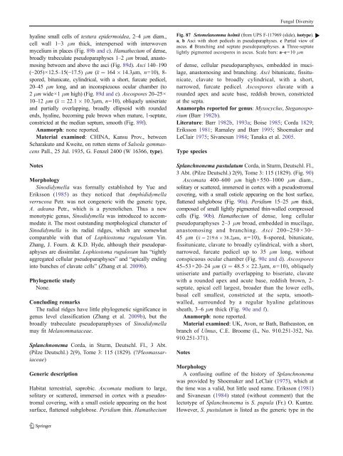
Fig. 87 Setomelanomma holmii (from UPS F-117969 (slide), isotype). b<br />
a, b Asci with short pedicels in pseudoparaphyses. c Partial view of<br />
ascus. d Branching and septate pseudoparaphyses. a Three-septate<br />
lightly pigmented ascospores in ascus. Scale bars: a–e=10 μm<br />
of dense, cellular pseudoparaphyses, embedded in mucilage,<br />
anastomosing and branching. Asci bitunicate, fissitunicate,<br />
clavate to broadly cylindrical, with a short,<br />
narrowed, furcate pedicel. Ascospores clavate with a<br />
rounded apex and acute base, reddish brown, constricted<br />
at the septa.<br />
Anamorphs reported for genus: Myxocyclus, Steganosporium<br />
(Barr 1982b).<br />
Literature: Barr 1982b, 1993a; Boise 1985; Corda 1829;<br />
Eriksson 1981; Ramaley and Barr 1995; Shoemaker and<br />
LeClair 1975; Sivanesan 1984; Tanaka et al. 2005.<br />
Type species<br />
Splanchnonema pustulatum Corda, in Sturm, Deutschl. Fl.,<br />
3 Abt. (Pilze Deutschl.) 2(9), Tome 3: 115 (1829). (Fig. 90)<br />
Ascomata 400–600 μm high×550–1000 μm diam.,<br />
solitary or scattered, immersed in cortex with a pseudostromal<br />
covering, with a small ostiole appearing on the host surface,<br />
flattened subglobose (Fig. 90a). Peridium 15–25 μm thick,<br />
composed of small lightly pigmented thin-walled compressed<br />
cells (Fig. 90b). Hamathecium of dense, long cellular<br />
pseudoparaphyses 2–3 μm broad, embedded in mucilage,<br />
anastomosing and branching. Asci 200–250×30–<br />
45 μm (x ¼ 219:6 38:2mm, n=10), 8-spored, bitunicate,<br />
fissitunicate, clavate to broadly cylindrical, with a short,<br />
narrowed, furcate pedicel up to 35 μm long, without<br />
conspicuous ocular chamber (Fig. 90c and d). Ascospores<br />
45–53×20–24 μm (x ¼ 48:5 22:3mm, n=10), obliquely<br />
uniseriate and partially overlapping to biseriate, clavate<br />
with a rounded apex and acute base, reddish brown, 2-<br />
septate, apical cell largest, broader than the lower cells,<br />
basal cell smallest, constricted at the septa, smoothwalled,<br />
surrounded by a regular hyaline gelatinous<br />
sheath, 3–6 μm thick (Fig. 90e and f).<br />
Anamorph: none reported.<br />
Material examined: UK, Avon, nr Bath, Batheaston, on<br />
branch of Ulmus, C.E. Broome (L, No. 910.251-352, No.<br />
910.251-371).<br />
Notes<br />
Morphology<br />
A confusing outline of the history of Splanchnonema<br />
was provided by Shoemaker and LeClair (1975), which at<br />
the time was a valid, but little used name. Eriksson (1981)<br />
and Sivanesan (1984) stated (without comment) that the<br />
lectotype of Splanchnonema is S. pupula (Fr.) O. Kuntze.<br />
However, S. pustulatum is listed as the generic type in the


