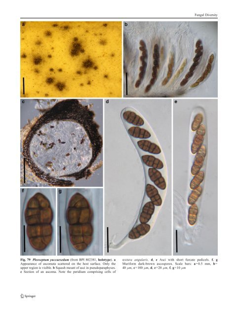
Fungal Diversity Fig. 79 Pleoseptum yuccaesedum (from BPI 802381, holotype). a Appearance of ascomata scattered on the host surface. Only the upper region is visible. b Squash mount of asci in pseudoparaphyses. c Section of an ascoma. Note the peridium comprising cells of textura angularis. d, e Asci with short furcate pedicels. f, g Muriform dark-brown ascospores. Scale bars: a=0.5 mm, b= 40 μm, c=100 μm, d, e=20 μm, f, g=10 μm
Fungal Diversity papillate, ostiolate. Peridium thin. Hamathecium of dense, cellular pseudoparaphyses. Asci 8-spored, bitunicate, fissitunicate, cylindrical to clavate, with furcate pedicel and small inconspicuous ocular chamber. Ascospores muriform, brown or pale brown, with or without sheath. Anamorphs reported for genus: Stemphylium (Simmons 1985). Literature: Barr 1981; Frisullo and Braun 1996; Kodsueb et al. 2006a; Luttrell1951; Wehmeyer1946, 1961, 1975; Zhang et al. 2009a. Type species Pleospora herbarum (Pers.) Rabenh., Klotzschii Herb. Viv. Mycol. 2: no. 547 (1854). (Fig. 80) ≡ Sphaeria herbarum Pers., Syn. meth. fung. (Göttingen) 1: 78 (1801). Ascomata 130–220 μm high×250–420 μm diam., scattered, or in small groups of 2–3, immersed, semiimmersed to erumpent, broadly to narrowly oblong and flattened, with flattened base not easily removed from the substrate, wall black, papillate, ostiolate (Fig. 80a and b). Peridium 30–50 μm thick on sides, thinner at the base, coriaceous, 2-layered, outer layer composed of one or two layers of heavily pigmented thick-walled cells of textura angularis, cells 5–10 μm diam., cell wall 2–4 μm thick, apex cells smaller and walls thicker, inner layer composed of hyaline thin-walled cells of textura angularis, 8–12 μm diam., wall hyaline, 0.5–1.5 μm thick (Fig. 80c). Hamathecium of dense, cellular pseudoparaphyses, 2–3 μm broad, filling the gaps between the asci. Asci 100–210× 27.5–30 μm (x ¼ 142:2 28:3mm, n=10), 8-spored, bitunicate, fissitunicate, broadly cylindrical to clavate, with a short, thick, furcate pedicel, 8-12(−20) μm long, with small inconspicuous ocular chamber (ca. 3 μm wide× 1 μm high) (Fig. 80d and e). Ascospores 28–38×12.5– 15 μm (x ¼ 33 14:5mm, n=10), ellipsoidal, straight or sometimes curved, with broadly rounded ends and upper hemispore slightly shorter and broader; spores usually divided by 3 A-transsepta, all 4 segments by longisepta andthenbyonestratumofB-transsepta(maturesporesas a rule with 7 transsepta, 3A+4B), yellowish brown, smooth; each hemispore with thick gelatinous sheath, the lower one with umbilicus (sheaths fused in mature spores) (Fig. 80f, g, h, i, j and k). Anamorph: Stemphyllium herbarum E. Simmons (Simmons 1985). Material examined: GERMANY, on stalks of Melilotusalla? at the bank of the Elbe in Konigstein, 1882 (E, Krieger 683); as Sphaeria herbarum Persoon Syn. fung. p. 78 (E, 81); as Sphaeria herbarum Fries, Scleromyceti Sueciae 38 (E, lectotype). Notes Morphology Pleospora was originally assigned within Sphaeriales. Subsequently, it was assigned within Pseudosphaeriales and <strong>Pleosporales</strong> (Wehmeyer 1961). Pleospora is a large group, which is widely distributed and associated with a wide range of species of monocotyledons as well as dicotyledons (Wehmeyer 1975). All species of Pleospora have muriform ascospores (Wehmeyer 1961, 1975). Pleospora has downward growing pseudoparaphyses within the ascomata of “Pleospora-type” development (Luttrell Univ. Mo. Stud. 1951), which subsequently served as a diagnostic character. However, only a limited number of species had detailed studies on this character (Wehmeyer 1961). The heterogeneous nature of Pleospora has been noted, and several subgenera have been erected, such as Scleroplea to include all “sclerotioid” species of Pleospora, Teichosporoides to accommodate species of Pleospora with immersed ascomata, Pleosphaeria for those having superficial and setose ascomata (Wehmeyer 1961). Similarly, Cucurbitaria, Fenestella and Montagnula are also separated as a section from Pleospora. Most of these subgenera are currently at genus rank. Phylogenetic study The polyphyletic nature of Pleospora is clear (Kodsueb et al. 2006a), and those that stain the woody substrate purple should be assigned to Amniculicolaceae (Zhang et al. 2009a). Concluding remarks As some Pleospora species have a wide range of host spectrum, especially on both monocotyledons and dicotyledons, it is highly possible they are cryptic species. Preussia Fuckel, Hedwigia 6: 175 (1867) [1869–70]. (Sporormiaceae) Generic description Habitat terrestrial, saprobic (on decaying fibers or coprophilous). Ascomata small- to medium-sized, cleistothecial or perithecial, solitary or scattered on substrate surface, globose, membraneous, black. Peridium thin, composed of thick-walled, poly-angular cells from the surface view. Pseudoparaphyses not observed. Asci (4-) 8-spored, bitunicate, clavate to broadly clavate, with a long and thin and furcate pedicel. Ascospores 3–6 seriate to uniseriate near the base, cylindrical with rounded ends, brown, septate, easily breaking into partspores, with germ slits in each cell. Anamorphs reported for genus: Phoma (von Arx 1973; Cain 1961; Malloch and Cain 1972). Literature: Ahmed and Cain 1972; Arenaletal.2005; von Arx 1973; von Arx and van der Aa 1987; Auerswald1866;
- Page 1 and 2:
Fungal Diversity DOI 10.1007/s13225
- Page 3 and 4:
Fungal Diversity Table 1 Major circ
- Page 5 and 6:
Fungal Diversity
- Page 7 and 8:
Fungal Diversity biocontrol agent o
- Page 9 and 10:
Fungal Diversity substrates and man
- Page 11 and 12:
Fungal Diversity 2. To investigate
- Page 13 and 14:
Fungal Diversity Table 3 (continued
- Page 15 and 16:
Fungal Diversity Table 3 (continued
- Page 17 and 18:
Fungal Diversity Table 3 (continued
- Page 19 and 20:
Fungal Diversity
- Page 21 and 22:
Fungal Diversity Fig. 2 Aigialus gr
- Page 23 and 24:
Fungal Diversity Fig. 3 Amniculicol
- Page 25 and 26:
Fungal Diversity Literature: Berkel
- Page 27 and 28:
Fungal Diversity Ascorhombispora L.
- Page 29 and 30:
Fungal Diversity
- Page 31 and 32:
Fungal Diversity Fig. 8 Astrosphaer
- Page 33 and 34:
Fungal Diversity Fig. 9 Asymmetrico
- Page 35 and 36:
Fungal Diversity Notes Morphology B
- Page 37 and 38:
Fungal Diversity Generic descriptio
- Page 39 and 40:
Fungal Diversity Anamorph: none rep
- Page 41 and 42:
Fungal Diversity Fig. 14 Bimuria no
- Page 43 and 44:
Fungal Diversity Fig. 15 Bricookea
- Page 45 and 46:
Fungal Diversity Fig. 16 Byssolophi
- Page 47 and 48:
Fungal Diversity Notes Morphology B
- Page 49 and 50:
Fungal Diversity the reaction of pe
- Page 51 and 52:
Fungal Diversity
- Page 53 and 54:
Fungal Diversity Fig. 21 Chaetomast
- Page 55 and 56:
Fungal Diversity
- Page 57 and 58:
Fungal Diversity Fig. 23 Cilioplea
- Page 59 and 60:
Fungal Diversity with one or two ve
- Page 61 and 62:
Fungal Diversity Moreau 1953; Munk
- Page 63 and 64:
Fungal Diversity Material examined:
- Page 65 and 66:
Fungal Diversity Fig. 28 Dothidotth
- Page 67 and 68:
Fungal Diversity Fig. 29 Dubitatio
- Page 69 and 70:
Fungal Diversity assigned Entodesmi
- Page 71 and 72:
Fungal Diversity fusoid to somewhat
- Page 73 and 74:
Fungal Diversity Fig. 33 Hadrospora
- Page 75 and 76:
Fungal Diversity Fig. 34 Halotthia
- Page 77 and 78:
Fungal Diversity Notes Morphology H
- Page 79 and 80:
Fungal Diversity some effused Hypox
- Page 81 and 82:
Fungal Diversity Fig. 38 Isthmospor
- Page 83 and 84:
Fungal Diversity Fig. 39 Kalmusia e
- Page 85 and 86:
Fungal Diversity ascospores were br
- Page 87 and 88:
Fungal Diversity furcate pedicel an
- Page 89 and 90:
Fungal Diversity Anamorph: none rep
- Page 91 and 92:
Fungal Diversity
- Page 93 and 94:
Fungal Diversity Material examined:
- Page 95 and 96:
Fungal Diversity Fig. 46 Lewia scro
- Page 97 and 98:
Fungal Diversity Fig. 47 Lichenopyr
- Page 99 and 100: Fungal Diversity Loculohypoxylon M.
- Page 101 and 102: Fungal Diversity cells small heavil
- Page 103 and 104: Fungal Diversity upper place, septa
- Page 105 and 106: Fungal Diversity
- Page 107 and 108: Fungal Diversity (CBS 627.86) was i
- Page 109 and 110: Fungal Diversity Fig. 54 Mamillisph
- Page 111 and 112: Fungal Diversity Fig. 55 Massarina
- Page 113 and 114: Fungal Diversity phaeria as a synon
- Page 115 and 116: Fungal Diversity 5-8 μm diam., ind
- Page 117 and 118: Fungal Diversity cell wall
- Page 119 and 120: Fungal Diversity Fig. 60 Mixtura sa
- Page 121 and 122: Fungal Diversity Fig. 61 Montagnula
- Page 123 and 124: Fungal Diversity spored, bitunicate
- Page 125 and 126: Fungal Diversity Fig. 64 Murispora
- Page 127 and 128: Fungal Diversity Type species Neoph
- Page 129 and 130: Fungal Diversity brown, 8-septate,
- Page 131 and 132: Fungal Diversity Fig. 68 Ohleria mo
- Page 133 and 134: Fungal Diversity Fig. 69 Ohleriella
- Page 135 and 136: Fungal Diversity Fig. 70 Ophiobolus
- Page 137 and 138: Fungal Diversity Type species Ostro
- Page 139 and 140: Fungal Diversity
- Page 141 and 142: Fungal Diversity (Shoemaker and Bab
- Page 143 and 144: Fungal Diversity ium thin, composed
- Page 145 and 146: Fungal Diversity Fig. 76 Platysporo
- Page 147 and 148: Fungal Diversity Fig. 77 1 Pleomass
- Page 149: Fungal Diversity Fig. 78 Pleophragm
- Page 153 and 154: Fungal Diversity Williams 1963; Mal
- Page 155 and 156: Fungal Diversity Generic descriptio
- Page 157 and 158: Fungal Diversity composed of one ce
- Page 159 and 160: Fungal Diversity Fig. 84 Saccharico
- Page 161 and 162: Fungal Diversity and nearly black a
- Page 163 and 164: Fungal Diversity dense, long trabec
- Page 165 and 166: Fungal Diversity
- Page 167 and 168: Fungal Diversity
- Page 169 and 170: Fungal Diversity Anamorphs reported
- Page 171 and 172: Fungal Diversity
- Page 173 and 174: Fungal Diversity
- Page 175 and 176: Fungal Diversity Fig. 94 Westerdyke
- Page 177 and 178: Fungal Diversity Fig. 95 Wettsteini
- Page 179 and 180: Fungal Diversity Fig. 96 Wilmia bra
- Page 181 and 182: Fungal Diversity Current name: Astr
- Page 183 and 184: Fungal Diversity spores are actuall
- Page 185 and 186: Fungal Diversity Fig. 100 Sporormie
- Page 187 and 188: Fungal Diversity
- Page 189 and 190: Fungal Diversity Fig. 102 Kriegerie
- Page 191 and 192: Fungal Diversity Phylogenetic study
- Page 193 and 194: Fungal Diversity Fig. 104 Zeuctomor
- Page 195 and 196: Fungal Diversity Fig. 105 Muroia ni
- Page 197 and 198: Fungal Diversity pseudoparenchymato
- Page 199 and 200: Fungal Diversity Eremodothis Arx, K
- Page 201 and 202:
Fungal Diversity Type species: Macr
- Page 203 and 204:
Fungal Diversity ascospores of Plat
- Page 205 and 206:
Fungal Diversity monoceras Alcorn n
- Page 207 and 208:
Fungal Diversity tomataceae, Melano
- Page 209 and 210:
Fungal Diversity Table 4 (continued
- Page 211 and 212:
Fungal Diversity 1987b). Based on a
- Page 213 and 214:
Fungal Diversity only do so under v
- Page 215 and 216:
Fungal Diversity Dennis RWG (1968)
- Page 217 and 218:
Fungal Diversity Kirk PM, Cannon PF
- Page 219 and 220:
Fungal Diversity Saccardo PA (1880)
- Page 221:
Fungal Diversity Winter G (1887) As