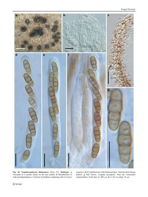
Fungal Diversity Fig. 66 Neophaeosphaeria filamentosa (from NY, holotype). a Ascomata as a circular cluster on the host surface. b Hamathecium of wide psuedoparaphyses. c Section of peridium comprising cells of textura angularis. d–f Cylindrical asci with thickened apex. Note the short furcate pedicel. g Pale brown, 3-septate ascospores. Note the verruculose ornamentation. Scale bars: a=200 μm, b, c=20 μm, d–g=10 μm
Fungal Diversity brown, 8-septate, the 4th upper cell broader than the others, smooth-walled, without sheath (Fig. 67e and f). Anamorph: none reported. Material examined: GERMANY, Dresdae, in herbarum caulibus emortuis perrara, exeunte majo, 1858 (BR 101945–95, holotype, asNodulosphaeria hirta). Notes Morphology The name Nodulosphaeria was first used by Rabenhorst (1858) but was considered as a synonym of Leptosphaeria for many years (Clements and Shear 1931). The name was reinstated by Holm (1957) and was represented by N. hirta, which was concurrently treated as a synonym of N. derasa (Berk. & Broome) L. Holm. The most outstanding morphological characters of Nodulosphaeria were considered to be apex of ascomata often covered with setae, ascospore with three or more transverse septa with a supramedian enlarged cell or elongated to a scolecospore, mostly with terminal appendages (Barr 1992a; Holm 1961; Shoemaker 1984b). The ascomata are usually immersed and the peridium comprises a few layers of brown, relatively thin-walled cells of textura angularis and textura prismatica similar to those of Phaeosphaeria. Thus, Nodulosphaeria is likely to be a member of Phaeosphaeriaceae. However,thisneedstobe confirmed by molecular analysis. The boundary between Nodulosphaeria and Ophiobolus is not clear-cut, and the circumscriptions of them usually depend on the viewpoint of different mycologists. For instance, Shoemaker (1976) has assigned some Nodulosphaeria species such as N. erythrospora, N. fruticum, N. mathieui and N. megalosporus to Ophiobolus. Subsequently, more species were added to Nodulosphaeria (Barr 1992a; Shoemaker 1984b; Shoemaker and Babcock 1987). Currently, more than 60 names are included in Nodulosphaeria (http://www.mycobank.org/, 06/ 2010). Phylogenetic study None. Concluding remarks All species included in Nodulosphaeria have an inflated ascospore cell as mentioned above. However, it is likely that this character would have evolved more than once as it is probably an adaption for ascospore ejection from the ascus (Shoemaker 1976). It occurs in Ophiobolus species and the ascomata of these species are quite dissimilar to Nodulosphaeria species and their exclusion from Nodulosphaeria seems warranted. When considering whether a species belongs in Nodulosphaeria, one must also consider the ascomata and peridium structure until DNA sequences are available. Ohleria Fuckel, Fungi rhenani exsic.: no. 2173 (1868). (Melanommataceae) Generic description Habitat terrestrial, saprobic. Ascomata small to medium size, solitary, scattered, or in small groups, erumpent to nearly superficial, papillate, ostiolate. Peridium thin, thicker at the apex, 1-layered. Hamathecium of dense, long trabeculate pseudoparaphyses. Asci 8-spored, bitunicate, fissitunicate, cylindrical, with a short pedicel. Ascospore brown to reddish brown, broadly to narrowly fusoid, 3-septate, easily separating into two parts at the primary septum. Anamorphs reported for genus: Monodictys (Samuels 1980). Literature: Barr 1990b; Clements and Shear 1931; Patel et al. 1997; Samuels 1980. Type species Ohleria modesta Fuckel, Fungi rhenani exsic. (1868) (Fig.68) Ascomata 214–357 μm high×285–400 μm diam., solitary, scattered, or in small groups of 2–3, erumpent to nearly superficial, coriaceous, with basal wall remaining immersed in host tissue, broadly or narrowly conical, with a flattened base not easily removed from the substrate, black; apex with a conical protruding papilla and an often porelike ostiole (Fig. 68a). Peridium 22–53 μm thick laterally, thicker at the apex, 1-layered, composed of heavily pigmented thick-walled cells of textura angularis, cells to 7 μm diam., cell wall 1.5–3 μm thick, apex cells smaller and walls thicker, base cells walls thinner (Fig. 68b). Hamathecium of dense, long trabeculate pseudoparaphyses 1–2 μm broad, septate, branching and anastomosing (Fig. 68c). Asci 90– 130×(5.5-)7–10 μm (x ¼ 107:3 8mm, n=10), 8-spored, with a short pedicel up to 20 μm long, bitunicate, fissitunicate, cylindrical, with a small ocular chamber (to 1.5 μm wide×1.5 μm high) (Fig. 68c, d and e). Ascospore 15–22×4–5 μm (x ¼ 20 4:4mm, n=10), biseriate near the top and uniseriate at the base, broadly fusoid to fusoid with broadly to narrowly rounded ends, brown to reddish brown, 3-septum, deeply constricted at the median septum and breaking into two conical partspores, no constriction at the secondary septum, smooth (Fig. 68d and e). Anamorph: none reported. Material examined: GERMANY, on decorticated, decaying roots of Fagus sylvatica, very rare, collected in autumn (G: F. rh. 2173, isotype). Notes Morphology Ohleria is characterized by its subglobose to conic ascomata, produced on decorticated woody substrates, as