

Pleosporales - CBS - KNAW
Pleosporales - CBS - KNAW
Pleosporales - CBS - KNAW

You also want an ePaper? Increase the reach of your titles
YUMPU automatically turns print PDFs into web optimized ePapers that Google loves.
Fungal Diversity<br />
ing between and above the asci (Fig. 92d, e and f). Asci<br />
100–145×15–17 μm (x ¼ 118 15:5mm, n=10), 8-spored,<br />
bitunicate, fissitunicate, cylindro-clavate, with a short,<br />
thick, furcate pedicel which is 12–30 μm long, with a<br />
truncate ocular chamber (Fig. 92d, e, f, g and i). Ascospores<br />
27.5–32.5×7.5–8.5 μm (x ¼ 29:5 8mm, n=10), biseriate<br />
to uniseriate near the base, fusoid with broadly to narrowly<br />
rounded ends, dark brown, 1–3-septate, secondary septum<br />
forming late or often absent, constricted at the median<br />
septum, the upper cell often shorter and broader than the<br />
lower one, smooth to finely verruculose, containing<br />
refractive globules (Fig. 92j and k).<br />
Anamorph: Only hyphopodia-like structures (or conidia?)<br />
observed (Zhang et al. 2008a).<br />
Colonies (of epitype) reaching 5 cm diam. after 20 days<br />
growth on MEA at 25°C, raised, woolly, deep grey, with<br />
irregular to rhizoidal margin, reverse darkened. Hyphopodialike<br />
structures (or conidia?) produced after 6 months, hyaline<br />
to pale brown, lobed, 4–4.5(−5) μm long and 3–3.5 μm diam.<br />
Material examined: EUROPE, Upsala, on decaying<br />
wood, designated by Boise (1985), (L-Pers 910269–172,<br />
as Sphaeria pertusa Pers., neotype); FRANCE, Deux<br />
Sèvres, Sansais, Le Vanneau, Les Grandes Mottines,<br />
swamp, on bark of a dead stump of Fraxinus excelsior,<br />
25 Apr. 2004, J. Fournier (IFRD 2002, epitype); Haute<br />
Garonne, Avignonet, Canal du Midi, on submerged wood<br />
of Platanus in a canal, 23 Nov. 2006, Michel Delpont, det.<br />
J. Fournier (IFRD2003).<br />
Notes<br />
Morphology<br />
Trematosphaeria was formally established in ‘Rhenish<br />
fungi’ by Fuckel (1870) based on the broadly pertuse<br />
ascomata, and Fries (1823) assigned it under Ascomycetes,<br />
Pyrenomycetes, Lophiostomataceae. Subsequently, Winter<br />
(1885) placed Trematosphaeria in Amphisphaeriaceae.<br />
Berlese (1890), however, treated Trematosphaeria as a<br />
synonym of Melanomma (Melanommataceae). After establishment<br />
of Loculoascomycetes (Luttrell 1955), Trematosphaeria<br />
was assigned to Pleosporaceae (Loculoascomycetes,<br />
<strong>Pleosporales</strong>) (Holm1957), and this was followed by von<br />
Arx and Müller (1975). Trematosphaeria was assigned to<br />
Melanommataceae by Barr (1979a), and this has been<br />
widely followed (Eriksson 2006; Kirketal.2001; Lumbsch<br />
and Huhndorf 2007).<br />
Trematosphaeria pertusa, the lectotype species of Trematosphaeria<br />
(Clements and Shear 1931), is characterized by<br />
having semi-immersed to erumpent ascomata, filamentous<br />
pseudoparaphyses, cylindro-clavate asci, fusoid, 1-septate<br />
reddish brown to dark brown ascospores (Zhang et al.<br />
2008a). All of these characters are quite different from those<br />
of Melanomma, the familial type of Melanommataceae.<br />
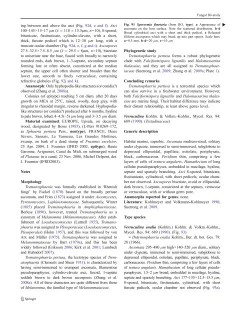
Fig. 91 Sporormia fimetaria (from RO, type). a Appearance of<br />
ascomata on the host surface. Note the scattered distribution. b–d<br />
Broad cylindrical asci with a short and thick pedicel. e Released<br />
filiform ascospores which may break up into part spores. Scale bars:<br />
a=0.5 mm, b–d=20 μm, e=10 μm<br />
Phylogenetic study<br />
Trematosphaeria pertusa forms a robust phylogenetic<br />
clade with Falciformispora lignatilis and Halomassarina<br />
thalassiae, and they are all assigned to Trematosphaeriaceae<br />
(Suetrong et al. 2009; Zhang et al. 2009a; Plate 1).<br />
Concluding remarks<br />
Trematosphaeria pertusa is a terrestrial species which<br />
can also survive in a freshwater environment. However,<br />
both Falciformispora lignatilis and Halomassarina thalassiae<br />
are marine fungi. Their habitat difference may indicate<br />
their distant relationship, at least above genus level.<br />
Verruculina Kohlm. & Volkm.-Kohlm., Mycol. Res. 94:<br />
689 (1990). (Testudinaceae)<br />
Generic description<br />
Habitat marine, saprobic. Ascomata medium-sized, solitary<br />
under clypeate, immersed to semi-immersed, subglobose to<br />
depressed ellipsoidal, papillate, ostiolate, periphysate,<br />
black, carbonaceous. Peridium thin, comprising a few<br />
layers of cells of textura angularis. Hamathecium of long<br />
cellular pseudoparaphyses, embedded in mucilage, hyaline,<br />
septate and sparsely branching. Asci 8-spored, bitunicate,<br />
fissitunicate, cylindrical, with short pedicels, ocular chamber<br />
not observed. Ascospores biseriate, ovoid or ellipsoidal,<br />
dark brown, 1-septate, constricted at the septum, verrucose<br />
or verruculose, with or without germ pore.<br />
Anamorphs reported for genus: none.<br />
Literature: Kohlmeyer and Volkmann-Kohlmeyer 1990;<br />
Suetrong et al. 2009.<br />
Type species<br />
Verruculina enalia (Kohlm.) Kohlm. & Volkm.-Kohlm.,<br />
Mycol. Res. 94: 689 (1990). (Fig. 93)<br />
≡ Didymosphaeria enalia Kohlm., Ber. dt. bot. Ges. 79:<br />
28 (1966).<br />
Ascomata 295–480 μm high×140–520 μm diam., solitary<br />
under clypeate, immersed to semi-immersed, subglobose to<br />
depressed ellipsoidal, ostiolate, papillate, periphysate, black,<br />
carbonaceous. Peridium thin, comprising a few layers of cells<br />
of textura angularis. Hamathecium of long cellular pseudoparaphyses,<br />
1.5–2 μm broad, embedded in mucilage, hyaline,<br />
septate and sparsely branching. Asci 177–135×12.5–15.5 μm,<br />
8-spored, bitunicate, fissitunicate, cylindrical, with short<br />
furcate pedicels, ocular chamber not observed (Fig. 93a).<br />
b


