

Pleosporales - CBS - KNAW
Pleosporales - CBS - KNAW
Pleosporales - CBS - KNAW

You also want an ePaper? Increase the reach of your titles
YUMPU automatically turns print PDFs into web optimized ePapers that Google loves.
Fungal Diversity<br />
dense, long trabeculate pseudoparaphyses, anastomosing<br />
and branching between the asci. Asci bitunicate, fissitunicate,<br />
cylindrical to cylindro-clavate, with a short furcate<br />
pedicel, with a big and truncate ocular chamber. Ascospores<br />
obliquely uniseriate and partially overlapping,<br />
narrowly fusoid to fusoid or broadly fusoid with tapering<br />
or narrowly rounded ends, hyaline to pale brown or<br />
brown, muriform.<br />
Anamorphs reported for genus: coelomycetous with<br />
muriform conidia (see Liu 2009).<br />
Literature: Cheng et al. 2004; Hino 1961; Kishi et al.<br />
1991; Liu 2009; Morakotkarn et al. 2008.<br />
Type species<br />
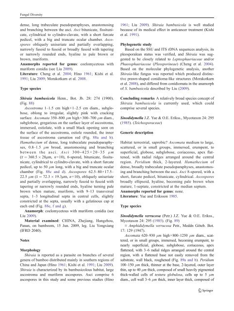
Shiraia bambusicola Henn., Bot. Jb. 28: 274 (1900).<br />
(Fig. 88)<br />
Ascostroma 1–1.5 cm high×1–2.5 cm diam., subglobose,<br />
oblong to irregular, slightly pink with cracking<br />
surface. Ascomata 350–800 μm high×300–700 μm diam.,<br />
subglobose, gregarious on the surface layer of ascostroma,<br />
immersed, ostiolate, with a small black opening seen on<br />
the surface of the ascostroma, ostiole rounded, the inner<br />
tissue of ascostroma carnation red (Fig. 88a and b).<br />
Hamathecium of dense, long trabeculate pseudoparaphyses,<br />
0.8–1.5 μm broad, anastomosing and branching<br />
between the asci. Asci 300– 425×20– 35 μm<br />
(x ¼ 360:5 28mm, n=10), 6-spored, bitunicate, fissitunicate,<br />
cylindrical to cylindro-clavate, with a short furcate<br />
pedicel, up to 50 μm long, with a big and truncate ocular<br />
chamber (Fig. 88c and d). Ascospores 62.5–80×17.5–<br />
22.5 μm (x ¼ 72:3 19:3mm, n=10), obliquely uniseriate<br />
and partially overlapping, narrowly fusoid to fusoid with<br />
tapering or narrowly rounded ends, hyaline turning pale<br />
brown when mature, muriform, with 9–13 transversal<br />
septa, 1–3 longitudinal septa in central cells, slightly<br />
constricted at the septa, usually with a gelatinous cap at<br />
each end (Fig. 88e, f and g).<br />
Anamorph: coelomycetous with muriform conidia (see<br />
Liu 2009).<br />
Material examined: CHINA, Zhejiang, Hangzhou,<br />
Panan, on bamboom, 15 Jun. 2009, leg. Liu Yongxiang<br />
(IFRD 2040).<br />
Notes<br />
Morphology<br />
Shiraia is reported as a parasite on branches of several<br />
genera of bamboo distributed mainly in southern regions of<br />
China and Japan (Hino 1961; Kishi et al. 1991; Liu 2009).<br />
Shiraia is characterized by its bambusicolous habitat, large<br />
ascostroma and muriform ascospores. Asci comprise 6<br />
ascospores in this study and some previous studies (Hino<br />
1961; Liu 2009). Shiraia bambusicola is well studied<br />
because of its medical effect in anticancer treatment (Kishi<br />
et al. 1991).<br />
Phylogenetic study<br />
Based on the SSU and ITS rDNA sequences analysis, its<br />
pleosporalean status was verified, and Shiraia was suggested<br />
to be closely related to Leptosphaeriaceae and/or<br />
Phaeosphaeriaceae (Pleosporineae) (Cheng et al. 2004).<br />
Based on the molecular phylogenetic analysis, another<br />
Shiraia-like fungus was reported which produced distinctive<br />
prawn-shaped conidioma-like structures (Morakotkarn<br />
et al. 2008), and differed from conidiomata in the anamorph<br />
of S. bambusicola described by Liu (2009).<br />
Concluding remarks A relatively broad species concept of<br />
Shiraia bambusicola is currently used, which could<br />
comprise several species.<br />
Sinodidymella J.Z. Yue & O.E. Erikss., Mycotaxon 24: 295<br />
(1985). (Teichosporaceae)<br />
Generic description<br />
Habitat terrestrial, saprobic? Ascomata medium to large,<br />
scattered, or in small groups, immersed, erumpent, to<br />
superficial, globose, subglobose, coriaceous, apex flattened,<br />
with radial ridges arranged around the central<br />
region. Peridium thick, 2-layered. Hamathecium of<br />
dense, broadly trabeculate pseudoparaphyses, anastomosing<br />
and branching between the asci. Asci 8-spored, with a<br />
short, furcate pedicel, bitunicate, cylindrical. Ascospores<br />
broadly ellipsoid, hyaline, becoming pale brown when<br />
mature, 1-septate, constricted at the median septum.<br />
Anamorphs reported for genus: none.<br />
Literature: Yue and Eriksson 1985.<br />
Type species<br />
Sinodidymella verrucosa (Petr.) J.Z. Yue & O.E. Erikss.,<br />
Mycotaxon 24: 295 (1985). (Fig. 89)<br />
≡ Amphididymella verrucosa Petr., Meddn Göteb. Bot.<br />
17: 129 (1947).<br />
Ascomata 620–930 μm high×800–1250 μm diam., scattered,<br />
or in small groups, immersed, becoming erumpent, to<br />
nearly superficial, globose, subglobose, coriaceous, apex<br />
flattened, with 3–6 radial ridges arranged around the central<br />
region, with a flattened base not easily removed from the<br />
substrate, wall black, roughened (Fig. 89a and b). Peridium<br />
100–150 μm thick, thinner at the base, 2-layered, outer layer<br />
thin, up to 40 μm thick, composed of small heavily pigmented<br />
thick-walled cells of textura globulosa, cells up to 5 μm<br />
diam., cell wall 3–6 μm thick, inner layer thick, composed of


