Fungal Diversity Anamorph: Pycnidia typical of Stagonospora (Sphaeropsidales), “scattered,arisingsinglybothonthehostandinpure culture, in culture generally surrounded by an envelope of mycelial hyphae, numerous, immersed on the host, but nearly superficial in culture, subglobose to slightly applanate, black, 150–250 μm diam., with a central slightly papillate ostiole, lacking a distinct neck; walls mainly 15–20 μm thick, composed of three to six layers of pseudoparenchymatous cells, the outermost layers dark brown and inner pale brown to hyaline cells somewhat compressed radially, very variable in size, cells of the outer layers mainly 7–12 μm long×4–6 μm wide in vertically section and 10–12 μm diam. in surface view, wall not or only slightly thicked near the ostiole. Conidiogenous cells lining the inner surface of the pycnidial cavity, holoblastic, minute and difficult to distinguish from the pseudoparenchymatous cells with which they are mixed, mammiform with a flattened apex, hyaline, smooth walled, about 4–6 μm tall and 4–6 μm wide. Conidia copiously produced, ellipsoid, with somewhat truncated ends, hyaline, smooth walled, (2-)3 septate, not or slightly constricted at the septa, often guttulate, rather thin walled, (21-)24–28(−34) μm×7–8.5(−11.5) μm” (from Kaiser et al. 1979). Material examined: KENYA, near Nairobi, on leaves of Saccharum officinarum L.; 24 Aug. 1977; leg. W.J. Kaiser (IMI 215888, holotype). Notes Morphology Saccharicola was separated from Leptosphaeria as a new genus based on its Stagonospora anamorph and its biotrophic habitat in leaves of sugar cane, and two species were included, i.e. Saccharicola bicolor and S. taiwanensis (J.M. Yen & C.C. Chi) O.E. Erikss. & D. Hawksw. (Eriksson and Hawksworth 2003). Saccharicola is characterized by its parasitic habitat on monocots, small ascomata, bitunicate asci, presence of pseudoparaphyses as well as its 3-septate ascospores (Eriksson and Hawksworth 2003). Phylogenetic study Based on the limited phylogenetic analysis of SSU sequences, Saccharicola is considered to be closely related to Massarina eburnea, the generic type of Massarina (Eriksson and Hawksworth 2003). Thus, Saccharicola was assigned to Massarinaceae, which includes Keissleriella, Massarina and Saccharicola (Eriksson and Hawksworth 2003). Concluding remarks Based on the parasitic habitat on monocots and its small ascomata and Stagonospora (or Cercospora? for S. taiwanensis, see Eriksson and Hawksworth 2003; Shoemaker and Babcock 1989b) anamorph, Saccharicola seems more similar to Pleosporineae. Further molecular study is needed for confirmation. Salsuginea K.D. Hyde, Bot. Mar. 34: 315 (1991). (<strong>Pleosporales</strong>, genera incertae sedis) Generic description Habitat marine, saprobic. Ascomata large, solitary, fusoid, conical or subglobose, with or without a flattened base, immersed under a darkened clypeus, papillate, ostiolate. Peridium thin, composed of round cells (in cross section) at sides, fusing at the top with the clypeus, thin at the base. Hamathecium of dense, long trabeculate pseudoparaphyses, anastomosing, embedded in mucilage. Asci 8-spored, bitunicate, fissitunicate, clavate to cylindro-clavate, pedunculate, with a large ocular chamber and conspicuous apical ring. Ascospores uniseriate, obovoid, brown to black, with hyaline apical germ pores, 1-septate, constricted at the septum, dark brown with paler apical cells, lacking sheath, smooth. Anamorphs reported for genus: none. Literature: Hyde 1991a; Suetrong et al. 2009. Type species Salsuginea ramicola K.D. Hyde, Bot. Mar. 34: 316 (1991). (Fig. 85) Ascomata 1040–2600 μm high×455–1430 μm diam., solitary, fusoid, conical or subglobose, with or without a flattened base, immersed under a darkened clypeus, papillate, ostiolate, ostiole rounded (Fig. 85a). Peridium up to 39 μm thick, composed of round cells (in cross section) at sides, fusing at the top with the clypeus, thin at the base (Fig. 85b). Hamathecium of dense, long trabeculate pseudoparaphyses, 1–2 μm broad, anastomosing, embedded in mucilage. Asci 440–512×29–34 μm, 8-spored, bitunicate, fissitunicate, clavate to cylindro-clavate, pedunculate, with a large ocular chamber and conspicuous apical ring (Fig. 85c and e). Ascospores 59–72×24–30 μm, uniseriate, obovoid, brown to black, with hyaline apical germ pores, 1-septate, constricted at the septum, dark brown with paler apical cells, lacking sheath, smooth (Fig. 85d and f). Anamorph: none reported. Material examined: THAILAND, Ranong mangrove, Aegiceras corniculatum (L.) Blanco., Oct. 1988, leg. & det. K.D. Hyde (BRIP 17102, holotype). Notes Morphology Salsuginea was introduced to accommodate the mangrove fungus, S. ramicola, which is characterized by large, immersed, ostiolate and papillate ascomata under a clypeus,
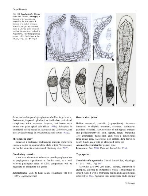
Fungal Diversity Fig. 84 Saccharicola bicolor (from IMI 215888, holotype). a Section of an ascomata immersed in the host tissue. b Section of a partial pycnidia. Note the phragmosporous conidia. c Clavate ascus with ocular chamber and short pedicel. d Ascospores. Note the pigmented central cell(s). Scale bars: a, b= 50 μm, c=20 μm, d=10 μm dense, trabeculate pseudoparaphyses embedded in gel matrix, fissitunicate, 8-spored, cylindrical asci with short pedicel and conspicuous apical apparatus, 1-septate, dark brown ascospores with paler apical cells (Hyde 1991a). Salsuginea is considered closely related to Helicascus and Caryospora, and they are all proposed to Melanommataceae (Hyde 1991a). Phylogenetic study Based on a multigene phylogenetic analysis, Salsuginea ramicola nested in a paraphyletic clade within <strong>Pleosporales</strong>; its familial status is undetermined (Suetrong et al. 2009). Concluding remarks It has been shown that trabeculate pseudoparaphyses has no phylogenetic significance at familial rank, so a well resolved phylogeny based on DNA comparisons will be necessary to categorize this genus. Semidelitschia Cain & Luck-Allen, Mycologia 61: 581 (1969). (Delitschiaceae) Generic description Habitat terrestrial, saprobic (coprophilous). Ascomata immersed to slightly erumpent, scattered, coriaceous, papillate, ostiolate. Hamathecium of non-typical trabeculate pseudoparaphyses, thin, septate, rarely branching. Asci cylindrical, pedicellate, each with a conspicuous large apical ring. Ascospores non-septate, dark brown to nearly black, each with an elongated germ slit. Anamorphs reported for genus: none. Literature: Barr 2000; Cain and Luck-Allen 1969. Type species Semidelitschia agasmatica Cain & Luck-Allen, Mycologia 61: 581 (1969). (Fig. 86) Ascomata 550–900 μm diam., solitary, immersed to erumpent, globose to subglobose, black, semicoriaceous, smooth-walled, with a protruding papilla and a conspicuous ostiole (Fig. 86a). Peridium thin, comprising multi-angular
- Page 1 and 2:
Fungal Diversity DOI 10.1007/s13225
- Page 3 and 4:
Fungal Diversity Table 1 Major circ
- Page 5 and 6:
Fungal Diversity
- Page 7 and 8:
Fungal Diversity biocontrol agent o
- Page 9 and 10:
Fungal Diversity substrates and man
- Page 11 and 12:
Fungal Diversity 2. To investigate
- Page 13 and 14:
Fungal Diversity Table 3 (continued
- Page 15 and 16:
Fungal Diversity Table 3 (continued
- Page 17 and 18:
Fungal Diversity Table 3 (continued
- Page 19 and 20:
Fungal Diversity
- Page 21 and 22:
Fungal Diversity Fig. 2 Aigialus gr
- Page 23 and 24:
Fungal Diversity Fig. 3 Amniculicol
- Page 25 and 26:
Fungal Diversity Literature: Berkel
- Page 27 and 28:
Fungal Diversity Ascorhombispora L.
- Page 29 and 30:
Fungal Diversity
- Page 31 and 32:
Fungal Diversity Fig. 8 Astrosphaer
- Page 33 and 34:
Fungal Diversity Fig. 9 Asymmetrico
- Page 35 and 36:
Fungal Diversity Notes Morphology B
- Page 37 and 38:
Fungal Diversity Generic descriptio
- Page 39 and 40:
Fungal Diversity Anamorph: none rep
- Page 41 and 42:
Fungal Diversity Fig. 14 Bimuria no
- Page 43 and 44:
Fungal Diversity Fig. 15 Bricookea
- Page 45 and 46:
Fungal Diversity Fig. 16 Byssolophi
- Page 47 and 48:
Fungal Diversity Notes Morphology B
- Page 49 and 50:
Fungal Diversity the reaction of pe
- Page 51 and 52:
Fungal Diversity
- Page 53 and 54:
Fungal Diversity Fig. 21 Chaetomast
- Page 55 and 56:
Fungal Diversity
- Page 57 and 58:
Fungal Diversity Fig. 23 Cilioplea
- Page 59 and 60:
Fungal Diversity with one or two ve
- Page 61 and 62:
Fungal Diversity Moreau 1953; Munk
- Page 63 and 64:
Fungal Diversity Material examined:
- Page 65 and 66:
Fungal Diversity Fig. 28 Dothidotth
- Page 67 and 68:
Fungal Diversity Fig. 29 Dubitatio
- Page 69 and 70:
Fungal Diversity assigned Entodesmi
- Page 71 and 72:
Fungal Diversity fusoid to somewhat
- Page 73 and 74:
Fungal Diversity Fig. 33 Hadrospora
- Page 75 and 76:
Fungal Diversity Fig. 34 Halotthia
- Page 77 and 78:
Fungal Diversity Notes Morphology H
- Page 79 and 80:
Fungal Diversity some effused Hypox
- Page 81 and 82:
Fungal Diversity Fig. 38 Isthmospor
- Page 83 and 84:
Fungal Diversity Fig. 39 Kalmusia e
- Page 85 and 86:
Fungal Diversity ascospores were br
- Page 87 and 88:
Fungal Diversity furcate pedicel an
- Page 89 and 90:
Fungal Diversity Anamorph: none rep
- Page 91 and 92:
Fungal Diversity
- Page 93 and 94:
Fungal Diversity Material examined:
- Page 95 and 96:
Fungal Diversity Fig. 46 Lewia scro
- Page 97 and 98:
Fungal Diversity Fig. 47 Lichenopyr
- Page 99 and 100:
Fungal Diversity Loculohypoxylon M.
- Page 101 and 102:
Fungal Diversity cells small heavil
- Page 103 and 104:
Fungal Diversity upper place, septa
- Page 105 and 106:
Fungal Diversity
- Page 107 and 108: Fungal Diversity (CBS 627.86) was i
- Page 109 and 110: Fungal Diversity Fig. 54 Mamillisph
- Page 111 and 112: Fungal Diversity Fig. 55 Massarina
- Page 113 and 114: Fungal Diversity phaeria as a synon
- Page 115 and 116: Fungal Diversity 5-8 μm diam., ind
- Page 117 and 118: Fungal Diversity cell wall
- Page 119 and 120: Fungal Diversity Fig. 60 Mixtura sa
- Page 121 and 122: Fungal Diversity Fig. 61 Montagnula
- Page 123 and 124: Fungal Diversity spored, bitunicate
- Page 125 and 126: Fungal Diversity Fig. 64 Murispora
- Page 127 and 128: Fungal Diversity Type species Neoph
- Page 129 and 130: Fungal Diversity brown, 8-septate,
- Page 131 and 132: Fungal Diversity Fig. 68 Ohleria mo
- Page 133 and 134: Fungal Diversity Fig. 69 Ohleriella
- Page 135 and 136: Fungal Diversity Fig. 70 Ophiobolus
- Page 137 and 138: Fungal Diversity Type species Ostro
- Page 139 and 140: Fungal Diversity
- Page 141 and 142: Fungal Diversity (Shoemaker and Bab
- Page 143 and 144: Fungal Diversity ium thin, composed
- Page 145 and 146: Fungal Diversity Fig. 76 Platysporo
- Page 147 and 148: Fungal Diversity Fig. 77 1 Pleomass
- Page 149 and 150: Fungal Diversity Fig. 78 Pleophragm
- Page 151 and 152: Fungal Diversity papillate, ostiola
- Page 153 and 154: Fungal Diversity Williams 1963; Mal
- Page 155 and 156: Fungal Diversity Generic descriptio
- Page 157: Fungal Diversity composed of one ce
- Page 161 and 162: Fungal Diversity and nearly black a
- Page 163 and 164: Fungal Diversity dense, long trabec
- Page 165 and 166: Fungal Diversity
- Page 167 and 168: Fungal Diversity
- Page 169 and 170: Fungal Diversity Anamorphs reported
- Page 171 and 172: Fungal Diversity
- Page 173 and 174: Fungal Diversity
- Page 175 and 176: Fungal Diversity Fig. 94 Westerdyke
- Page 177 and 178: Fungal Diversity Fig. 95 Wettsteini
- Page 179 and 180: Fungal Diversity Fig. 96 Wilmia bra
- Page 181 and 182: Fungal Diversity Current name: Astr
- Page 183 and 184: Fungal Diversity spores are actuall
- Page 185 and 186: Fungal Diversity Fig. 100 Sporormie
- Page 187 and 188: Fungal Diversity
- Page 189 and 190: Fungal Diversity Fig. 102 Kriegerie
- Page 191 and 192: Fungal Diversity Phylogenetic study
- Page 193 and 194: Fungal Diversity Fig. 104 Zeuctomor
- Page 195 and 196: Fungal Diversity Fig. 105 Muroia ni
- Page 197 and 198: Fungal Diversity pseudoparenchymato
- Page 199 and 200: Fungal Diversity Eremodothis Arx, K
- Page 201 and 202: Fungal Diversity Type species: Macr
- Page 203 and 204: Fungal Diversity ascospores of Plat
- Page 205 and 206: Fungal Diversity monoceras Alcorn n
- Page 207 and 208: Fungal Diversity tomataceae, Melano
- Page 209 and 210:
Fungal Diversity Table 4 (continued
- Page 211 and 212:
Fungal Diversity 1987b). Based on a
- Page 213 and 214:
Fungal Diversity only do so under v
- Page 215 and 216:
Fungal Diversity Dennis RWG (1968)
- Page 217 and 218:
Fungal Diversity Kirk PM, Cannon PF
- Page 219 and 220:
Fungal Diversity Saccardo PA (1880)
- Page 221:
Fungal Diversity Winter G (1887) As