Fungal Diversity Phylogenetic study None. Concluding remarks Its small-sized ascomata, broadly cylindrical to slightly obclavate asci with a short, thick, knob-like pedicel, as well as its monocotyledonous host preference point Metameris to the Phaeosphaeriaceae. But DNA comparisons are needed for confirmation. Mixtura O.E. Erikss. & J.Z. Yue, Mycotaxon 38: 203 (1990). (Phaeosphaeriaceae) Generic description Habitat terrestrial, parasitic. Ascomata small-sized, scattered or clustered on the leaf spots, immersed, erumpent, minutely papillate, ostiolate. Papilla slightly raised. Peridium thin, comprising one cell type of lightly pigmented thin-walled cells of textura angularis. Hamathecium of dense, filliform, septate, cellular pseudoparaphyses, 4– 6.3 μm broad, embedded in mucilage. Asci bitunicate, ovoid, with a very short stumpy pedicel. Ascospores fusoid to narrowly fusoid with broadly to narrowly rounded ends, curved, dark brown, multi-septate, distoseptate, with a germ pore at the lower end. Anamorphs reported for genus: none. Literature: Eriksson and Yue 1990. Type species Mixtura saginata (Syd.) O.E. Erikss. & J.Z. Yue, Mycotaxon 38: 203 (1990). (Fig. 60) ≡ Leptosphaeria saginata Syd., Annls mycol. 37: 376 (1939). Producing elongated yellow spots with brownish margins, leaf spots up to 45×3–5 mm, opposite side visible as a brownish spots (Fig. 60a). Ascomata 170–200 μm high×210– 280 μm diam., scattered on the lower side of the leaf, immersed, erumpent, breaking through the epidermis, minutely papillate. Papilla central, slightly raised, ostiolate, ostiole surrounded by a white margin (Fig. 60b). Peridium 22–34 μm wide, thicker at the apex, thinner at the base, comprising one cell type of lightly pigmented thin-walled cells of textura angularis, cells up to 6×8 μm diam., cell wall 0.5–1.2 μm thick, apex cells smaller and walls thicker (Fig. 60c). Hamathecium of dense, filliform, septate, cellular pseudoparaphyses, 4–6.3 μm broad, embedded in mucilage. Asci 80–128×41–53(−69) μm (x ¼ 100:9 52:8mm, n =10), 8-spored, bitunicate, fissitunicate dehiscence not observed, sac-like, with a very short stumpy pedicel and a small ocular chamber (Fig. 60d). Ascospores 86–94(−106)×20.5–23.5 μm (x ¼ 92:7 21:7mm, n=10), fasciculate, fusoid to narrowly fusoid, slightly curved, dark brown, 7-septate, distoseptate, with or without constriction at the primary septum, smoothwalled, with a germ pore at the lower end (Fig. 60e and f). Anamorph: none reported. Material examined: ECUADOR, Tungurahua, Hacienda San Antonio pr. Baños, Province, on the leaves of Chusqueae serrulatae Pilger., 9 Jan. 1938, H. Sydow. (S reg. nr F8934 type, F8935isolectotype, asLeptosphaeria saginata). Notes Morphology Mixtura was formally established by Eriksson and Yue (1990) as a monotypic genus represented by M. saginata basedonitsimmersedandthin-walledascomata, sparse, broad pseudoparaphyses, sac-like asci with a short pedicel and thick apex. Mixtura has a “mixture” of characters found in other pleosporalean genera. The peridium structure is comparable with Phaeosphaeria, the ascospores with Trematosphaeria and asci with Wettsteinina (Eriksson and Yue 1990).Accordingtothe structure of ascomata and hamathecium, Mixtura was provisionally assigned to Phaeosphaeriaceae (Eriksson and Yue 1990). Phylogenetic study None. Concluding remarks Morphologically, the sparse broad pseudoparaphyses and sac-like asci with a thick apical structure in Mixtura seem more comparable with the generic type of Teratosphaeria (T. fibrillose Syd. & P. Syd., Teratosphaeriaceae, Capnodiales, Dothideomycetidae) than that of Phaeosphaeria (P. oryzae). The heavily pigmented, multi-septate ascospores and the persistent pseudoparaphyses of Mixtura however, differ from those of Teratosphaeria. Thus, here we assign Mixtura under Teratosphaeriaceae as a distinct genus until phylogenetic work is carried out. Montagnula Berl., Icon. fung. (Abellini) 2: 68 (1896). (Montagnulaceae) Generic description Habitat terrestrial, saprobic. Ascomata small- to mediumsized, immersed to erumpent, gregarious or grouped, globose to subglobose, black. Hamathecium of dense, narrowly cellular, septate pseudoparaphyses. Asci bitunicate, fissitunicate, usually cylindro-clavate to clavate with a long pedicel. Ascospores oblong to narrowly oblong, straight or somewhat curved, reddish brown to dark yellowish brown, muriform or phragmosporous. Anamorphs reported for genus: Aschersonia (Hyde et al. 2011).
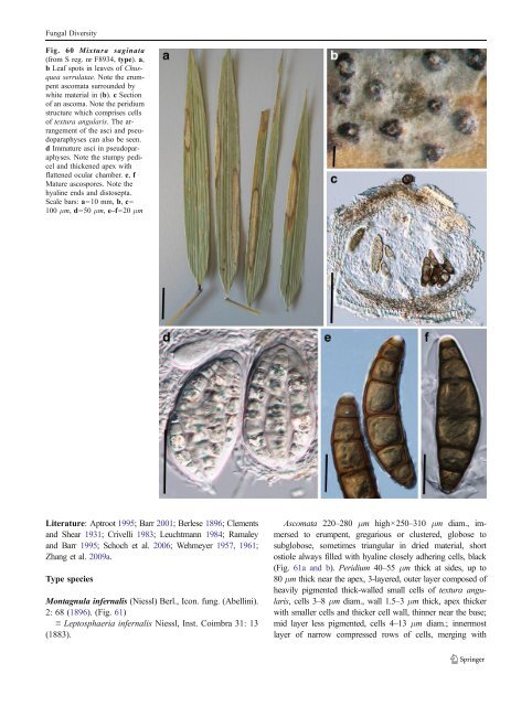
Fungal Diversity Fig. 60 Mixtura saginata (from S reg. nr F8934, type). a, b Leaf spots in leaves of Chusquea serrulatae. Note the erumpent ascomata surrounded by white material in (b). c Section of an ascoma. Note the peridium structure which comprises cells of textura angularis. The arrangement of the asci and pseudoparaphyses can also be seen. d Immature asci in pseudoparaphyses. Note the stumpy pedicel and thickened apex with flattened ocular chamber. e, f Mature ascospores. Note the hyaline ends and distosepta. Scale bars: a=10 mm, b, c= 100 μm, d=50 μm, e–f=20 μm Literature: Aptroot 1995; Barr2001; Berlese1896; Clements and Shear 1931; Crivelli1983; Leuchtmann 1984; Ramaley and Barr 1995; Schoch et al. 2006; Wehmeyer1957, 1961; Zhang et al. 2009a. Type species Montagnula infernalis (Niessl) Berl., Icon. fung. (Abellini). 2: 68 (1896). (Fig. 61) ≡ Leptosphaeria infernalis Niessl, Inst. Coimbra 31: 13 (1883). Ascomata 220–280 μm high×250–310 μm diam., immersed to erumpent, gregarious or clustered, globose to subglobose, sometimes triangular in dried material, short ostiole always filled with hyaline closely adhering cells, black (Fig. 61a and b). Peridium 40–55 μm thick at sides, up to 80 μm thick near the apex, 3-layered, outer layer composed of heavily pigmented thick-walled small cells of textura angularis, cells3–8 μm diam., wall 1.5–3 μm thick, apex thicker with smaller cells and thicker cell wall, thinner near the base; mid layer less pigmented, cells 4–13 μm diam.; innermost layer of narrow compressed rows of cells, merging with
- Page 1 and 2:
Fungal Diversity DOI 10.1007/s13225
- Page 3 and 4:
Fungal Diversity Table 1 Major circ
- Page 5 and 6:
Fungal Diversity
- Page 7 and 8:
Fungal Diversity biocontrol agent o
- Page 9 and 10:
Fungal Diversity substrates and man
- Page 11 and 12:
Fungal Diversity 2. To investigate
- Page 13 and 14:
Fungal Diversity Table 3 (continued
- Page 15 and 16:
Fungal Diversity Table 3 (continued
- Page 17 and 18:
Fungal Diversity Table 3 (continued
- Page 19 and 20:
Fungal Diversity
- Page 21 and 22:
Fungal Diversity Fig. 2 Aigialus gr
- Page 23 and 24:
Fungal Diversity Fig. 3 Amniculicol
- Page 25 and 26:
Fungal Diversity Literature: Berkel
- Page 27 and 28:
Fungal Diversity Ascorhombispora L.
- Page 29 and 30:
Fungal Diversity
- Page 31 and 32:
Fungal Diversity Fig. 8 Astrosphaer
- Page 33 and 34:
Fungal Diversity Fig. 9 Asymmetrico
- Page 35 and 36:
Fungal Diversity Notes Morphology B
- Page 37 and 38:
Fungal Diversity Generic descriptio
- Page 39 and 40:
Fungal Diversity Anamorph: none rep
- Page 41 and 42:
Fungal Diversity Fig. 14 Bimuria no
- Page 43 and 44:
Fungal Diversity Fig. 15 Bricookea
- Page 45 and 46:
Fungal Diversity Fig. 16 Byssolophi
- Page 47 and 48:
Fungal Diversity Notes Morphology B
- Page 49 and 50:
Fungal Diversity the reaction of pe
- Page 51 and 52:
Fungal Diversity
- Page 53 and 54:
Fungal Diversity Fig. 21 Chaetomast
- Page 55 and 56:
Fungal Diversity
- Page 57 and 58:
Fungal Diversity Fig. 23 Cilioplea
- Page 59 and 60:
Fungal Diversity with one or two ve
- Page 61 and 62:
Fungal Diversity Moreau 1953; Munk
- Page 63 and 64:
Fungal Diversity Material examined:
- Page 65 and 66:
Fungal Diversity Fig. 28 Dothidotth
- Page 67 and 68: Fungal Diversity Fig. 29 Dubitatio
- Page 69 and 70: Fungal Diversity assigned Entodesmi
- Page 71 and 72: Fungal Diversity fusoid to somewhat
- Page 73 and 74: Fungal Diversity Fig. 33 Hadrospora
- Page 75 and 76: Fungal Diversity Fig. 34 Halotthia
- Page 77 and 78: Fungal Diversity Notes Morphology H
- Page 79 and 80: Fungal Diversity some effused Hypox
- Page 81 and 82: Fungal Diversity Fig. 38 Isthmospor
- Page 83 and 84: Fungal Diversity Fig. 39 Kalmusia e
- Page 85 and 86: Fungal Diversity ascospores were br
- Page 87 and 88: Fungal Diversity furcate pedicel an
- Page 89 and 90: Fungal Diversity Anamorph: none rep
- Page 91 and 92: Fungal Diversity
- Page 93 and 94: Fungal Diversity Material examined:
- Page 95 and 96: Fungal Diversity Fig. 46 Lewia scro
- Page 97 and 98: Fungal Diversity Fig. 47 Lichenopyr
- Page 99 and 100: Fungal Diversity Loculohypoxylon M.
- Page 101 and 102: Fungal Diversity cells small heavil
- Page 103 and 104: Fungal Diversity upper place, septa
- Page 105 and 106: Fungal Diversity
- Page 107 and 108: Fungal Diversity (CBS 627.86) was i
- Page 109 and 110: Fungal Diversity Fig. 54 Mamillisph
- Page 111 and 112: Fungal Diversity Fig. 55 Massarina
- Page 113 and 114: Fungal Diversity phaeria as a synon
- Page 115 and 116: Fungal Diversity 5-8 μm diam., ind
- Page 117: Fungal Diversity cell wall
- Page 121 and 122: Fungal Diversity Fig. 61 Montagnula
- Page 123 and 124: Fungal Diversity spored, bitunicate
- Page 125 and 126: Fungal Diversity Fig. 64 Murispora
- Page 127 and 128: Fungal Diversity Type species Neoph
- Page 129 and 130: Fungal Diversity brown, 8-septate,
- Page 131 and 132: Fungal Diversity Fig. 68 Ohleria mo
- Page 133 and 134: Fungal Diversity Fig. 69 Ohleriella
- Page 135 and 136: Fungal Diversity Fig. 70 Ophiobolus
- Page 137 and 138: Fungal Diversity Type species Ostro
- Page 139 and 140: Fungal Diversity
- Page 141 and 142: Fungal Diversity (Shoemaker and Bab
- Page 143 and 144: Fungal Diversity ium thin, composed
- Page 145 and 146: Fungal Diversity Fig. 76 Platysporo
- Page 147 and 148: Fungal Diversity Fig. 77 1 Pleomass
- Page 149 and 150: Fungal Diversity Fig. 78 Pleophragm
- Page 151 and 152: Fungal Diversity papillate, ostiola
- Page 153 and 154: Fungal Diversity Williams 1963; Mal
- Page 155 and 156: Fungal Diversity Generic descriptio
- Page 157 and 158: Fungal Diversity composed of one ce
- Page 159 and 160: Fungal Diversity Fig. 84 Saccharico
- Page 161 and 162: Fungal Diversity and nearly black a
- Page 163 and 164: Fungal Diversity dense, long trabec
- Page 165 and 166: Fungal Diversity
- Page 167 and 168: Fungal Diversity
- Page 169 and 170:
Fungal Diversity Anamorphs reported
- Page 171 and 172:
Fungal Diversity
- Page 173 and 174:
Fungal Diversity
- Page 175 and 176:
Fungal Diversity Fig. 94 Westerdyke
- Page 177 and 178:
Fungal Diversity Fig. 95 Wettsteini
- Page 179 and 180:
Fungal Diversity Fig. 96 Wilmia bra
- Page 181 and 182:
Fungal Diversity Current name: Astr
- Page 183 and 184:
Fungal Diversity spores are actuall
- Page 185 and 186:
Fungal Diversity Fig. 100 Sporormie
- Page 187 and 188:
Fungal Diversity
- Page 189 and 190:
Fungal Diversity Fig. 102 Kriegerie
- Page 191 and 192:
Fungal Diversity Phylogenetic study
- Page 193 and 194:
Fungal Diversity Fig. 104 Zeuctomor
- Page 195 and 196:
Fungal Diversity Fig. 105 Muroia ni
- Page 197 and 198:
Fungal Diversity pseudoparenchymato
- Page 199 and 200:
Fungal Diversity Eremodothis Arx, K
- Page 201 and 202:
Fungal Diversity Type species: Macr
- Page 203 and 204:
Fungal Diversity ascospores of Plat
- Page 205 and 206:
Fungal Diversity monoceras Alcorn n
- Page 207 and 208:
Fungal Diversity tomataceae, Melano
- Page 209 and 210:
Fungal Diversity Table 4 (continued
- Page 211 and 212:
Fungal Diversity 1987b). Based on a
- Page 213 and 214:
Fungal Diversity only do so under v
- Page 215 and 216:
Fungal Diversity Dennis RWG (1968)
- Page 217 and 218:
Fungal Diversity Kirk PM, Cannon PF
- Page 219 and 220:
Fungal Diversity Saccardo PA (1880)
- Page 221:
Fungal Diversity Winter G (1887) As