

Pleosporales - CBS - KNAW
Pleosporales - CBS - KNAW
Pleosporales - CBS - KNAW

You also want an ePaper? Increase the reach of your titles
YUMPU automatically turns print PDFs into web optimized ePapers that Google loves.
Fungal Diversity<br />
Ascospores 16.5–23×7.5–10 μm, biseriate, ovoid or ellipsoidal,<br />
dark brown, 1-septate, constricted at the septum, verrucose<br />
or verruculose, with or without germ pore (Fig. 93b).<br />
Anamorph: none reported.<br />
Material examined: SEYCHELLES, Victoria, on submerged<br />
branch of Rhizophora mangle L., Mar. 2004, K.D.<br />
Hyde (KDH 2137, slide).<br />
Notes<br />
Morphology<br />
Verruculina was introduced to accommodate an obligate<br />
marine species, i.e. Verruculina enalia (Kohlmeyer<br />
and Volkmann-Kohlmeyer 1990). Verruculina is characterized<br />
by immersed, clypeate, carbonaceous, ostiolate and<br />
papillate ascomata. The peridium is composed of cells of<br />
textura angularis. Pseudoparaphyses are trabeculate and<br />
embedded in mucilage. Asci are 8-spored, cylindrical with<br />
short pedicels and ocular chamber, and ascospores are<br />
ellipsoidal, 1-septate, dark brown, verrucose or verruculose.<br />
The partly or completely immersed clypeate ascomata<br />
of V. enalia is comparable with those of<br />
Didymosphaeria futilis, but it differs from the later by<br />
the dark peridium, gelatinous matrix around the pseudoparaphyses,<br />
stipitate asci with an ocular chamber, and the<br />
verruculose ascospores (Kohlmeyer and Volkmann-<br />
Kohlmeyer 1990).<br />
Phylogenetic study<br />
Based on multigene phylogenetic analysis, Verruculina<br />
enalia nested within Testudinaceae (Suetrong et al. 2009).<br />
Thus, its familial placement seems clarified.<br />
Concluding remarks<br />
None.<br />
Westerdykella Stolk, Trans. Br. Mycol. Soc. 38: 422<br />
(1955). (Sporormiaceae)<br />
Generic description<br />
Habitat terrestrial, saprobic (coprophilous). Ascomata<br />
small, scattered on the upper layer of the culture medium,<br />
wall black. Peridium thin, composed of one layer of cells of<br />
polygonal, dark brown, thick-walled cells. Hamathecium<br />
not observed. Asci 32-spored, bitunicate nature undetermined,<br />
fissitunicate dehiscence not observed, subglobose to<br />
ellipsoid, arranged in the centre of the ascomata, with or<br />
without a short pedicel. Ascospores globose, brown, 1-celled,<br />
without germ pore.<br />
Anamorphs reported for genus: Phoma-like (von Arx 1974).<br />
Literature: von Arx 1973, 1981; Kruys et al. 2006; Kruys<br />
and Wedin 2009; Stolk 1955a.<br />
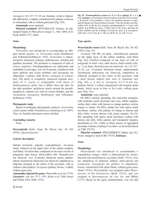
Fig. 92 Trematosphaeria pertusa (a, d, f–i from epitype, b, c, e, j b<br />
from neotype). a Ascomata on the host surface. b Section of an ascoma.<br />
c, h Section of the peridium. c shows the peridium structure at sides,<br />
and h indicates the basal peridium structure. Note the hyaline and thinwalled<br />
cells in (h). d Asci amongst pseudoparaphyses. e Ascus with<br />
pedicle. f, g Dehiscent ascus. i Upper part of the ascus, showing the<br />
ocular chamber and the mucilage covering the apex. j, k Ascospores.<br />
Scale bars: a=0.5 mm, b, c=100 μm, d–h=20 μm, i–k=10 μm<br />
Type species<br />
Westerdykella ornata Stolk, Trans. Br. Mycol. Soc. 38: 422<br />
(1955). (Fig. 94)<br />
Ascomata 100–300 μm diam., cleistothecoid, scattered<br />
on the upper layer of the culture medium, wall black<br />
(Fig. 94a). Peridium composed of one layer of cells of<br />
polygonal in front view, dark brown, thick-walled cells,<br />
ca. 5μm diam. Hamathecium not observed. Asci 25–32×<br />
16–22 μm, 32-spored, bitunicate nature undetermined,<br />
fissitunicate dehiscence not observed, subglobose to<br />
ellipsoid, arranged in the centre of the ascomata, with<br />
a short furcate pedicel best seen in immature asci<br />
(Fig. 94b, c, d and f). Ascospores 6.2–7×6–6.8 μm,<br />
globose, brown, 1-celled, ornamented with irregular spiral<br />
bands, which occur in four to five coils, without germ<br />
pore (Fig. 94e).<br />
Anamorph: none reported.<br />
On MEA colonies spreading, but somewhat erumpent,<br />
with moderate aerial mycelium and even, lobate margins;<br />
surface dirty white with luteous to orange patches; reverse<br />
orange to sienna. On PDA similar but with sparse aerial<br />
mycelium; surface with patches of orange to luteous and<br />
dirty white; reverse luteous with cream margins. On OA<br />
flat, spreading with sparse aerial mycelium; surface with<br />
luteous and dirty white patches and transparent margins;<br />
sporulating on OA, visible as black masses of aggregated<br />
ascomata; colonies reaching 4 cm diam. on all media (based<br />
on <strong>CBS</strong> 379.55).<br />
Material examined: MOZAMBIQUE, Inhaca, leg. H.J.<br />
Swart, mangrove mud (<strong>CBS</strong> 379.55, holotype).<br />
Notes<br />
Morphology<br />
Westerdykella was introduced to accommodate a<br />
coprophilous fungus, which is characterized by cleistothecioid<br />
and membraneous ascomata (Stolk 1955a). Asci<br />
are subglobose to ellipsoid, stalked, many-spored and<br />
evanescent. Ascospores are globose to subglobose,<br />
brown, ornamented with spiral bands, without germ pores<br />
(Stolk 1955a). Westerdykella was assigned under Phaeosporeae<br />
of the Eurotiaceae (Stolk 1955a), and was<br />
assigned to Sporormiaceae by von Arx and Müller<br />
(1975). Based on the spore ornamentation, von Arx and


