

Pleosporales - CBS - KNAW
Pleosporales - CBS - KNAW
Pleosporales - CBS - KNAW

You also want an ePaper? Increase the reach of your titles
YUMPU automatically turns print PDFs into web optimized ePapers that Google loves.
Fungal Diversity<br />
Anamorphs reported for genus: none.<br />
Literature: Ahmed and Asad 1968; Ahmed and Cain 1972;<br />
Kirschstein 1944; de Notaris 1849.<br />
Type species<br />
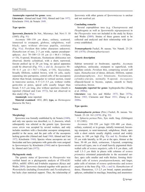
Sporormia fimetaria De Not., Micromyc. Ital. Novi 5: 10<br />
(1845). (Fig. 91)<br />
Ascomata 100–150 μm diam., solitary, scattered,<br />
immersed to erumpent, globose, subglobose, wall<br />
black; apex without obvious papilla, ostiolate<br />
(Fig. 91a). Peridium thin (other characters unknown).<br />
Hamathecium of rare, 2–3 μm wide, septate pseudoparaphyses.<br />
Asci 70–100×13–18 μm (x ¼ 86:4 14:9mm,<br />
n=10), 8-spored, bitunicate, fissitunicate dehiscence not<br />
observed, shortly cylindrical, with a short, narrowed,<br />
furcate pedicel up to 20 μm long, no apical apparatus<br />
could be observed (Fig. 91b, c and d). Ascospores 50–<br />
58×4–5 μm (x ¼ 54:7 4:8mm, n =10), fasciculate,<br />
broadly filliform, reddish brown, with 16 cells, easily<br />
separating into partspores, central cells of the ascospores<br />
shorter than broad, rectangular in vertical section, round<br />
in transverse section, 4–5×2.5–3.5 μm, without visible<br />
germ-slits or pores, apical cells usually longer than<br />
broad, 5–6.5 μm long, also without apertures (sheath is<br />
reported (Ahmed and Cain 1972), but not observed in<br />
this study) (Fig. 91e).<br />
Anamorph: none reported.<br />
Material examined: 1832, (RO, type, as Hormospora<br />
fimetaria De Not.).<br />
Notes<br />
Morphology<br />
Sporormia was formally established by de Notaris (1849),<br />
and only one species was described, i.e. S. fimetaria, which<br />
subsequently was selected as the generic type. Sporormia<br />
sensu stricto was accepted by several workers, and only<br />
includes members with a fasciculate ascospore arrangement,<br />
parallel to the ascus, and the part cells of the ascospores<br />
lacking germ-slits (Ahmed and Asad 1968; Ahmed and Cain<br />
1972; Kirschstein 1944). Species whose ascospores are not<br />
fasciculate and have partspores with germ-slits were assigned<br />
to Sporormiopsis by Kirschstein (1944) andtoSporormiella<br />
by Ahmed and Cain (1972).<br />
Phylogenetic study<br />
The generic status of Sporormia in <strong>Pleosporales</strong> was<br />
verified based on a phylogenetic analysis of ITS-nLSU<br />
rDNA, mtSSU rDNA and ß-tubulin sequences (Kruys and<br />
Wedin 2009). Sporormia clustered together with species of<br />
Westerdykella (including Eremodothis and Pycnidiophora),<br />
but lacks clear statistical support. Thus, the relationship of<br />
Sporormia with other genera of Sporormiaceae is unclear<br />
and not resolved yet.<br />
Concluding remarks<br />
Several coprophilous taxa (e.g. Chaetopreussia and<br />
Pleophragmia as well as Sporormiella nigropurpurea) in<br />
the <strong>Pleosporales</strong> were not included in the study by Kruys<br />
and Wedin (2009). Strains of these genera need to be<br />
collected and analyzed and their relationship with Sporormia<br />
established.<br />
Trematosphaeria Fuckel, Jb. nassau. Ver. Naturk. 23–24:<br />
161 (1870). (Trematosphaeriaceae)<br />
Generic description<br />
Habitat terrestrial or freshwater, saprobic. Ascomata<br />
subglobose, unilocular, erumpent to superficial, with<br />
papillate ostiole. Peridium thin, comprising several cell<br />
types. Hamathecium of dense, delicate, filliform, septate<br />
pseudoparaphyses. Asci bitunicate, fissitunicate,<br />
cylindro-clavate, normally 8-spored. Ascospores<br />
ellipsoid-fusoid to biconic, septate, smooth to finely<br />
verruculose, brown.<br />
Anamorphs reported for genus: hyphopodia-like (Zhang<br />
et al. 2008a).<br />
Literature: von Arx and Müller 1975; Barr 1979a;<br />
Boise 1985; Clements and Shear 1931; Zhang et al.<br />
2008a.<br />
Type species<br />
Trematosphaeria pertusa (Pers.) Fuckel, Jb. nassau. Ver.<br />
Naturk. 23–24: 161 (1870). (Fig. 92)<br />
≡ Sphaeria pertusa Pers., Syn. meth. fung. (Göttingen)<br />
1: 83 (1801).<br />
Ascomata 350–550 μm high×320–480 μm diam.,<br />
solitary, scattered, or in groups, initially immersed, becoming<br />
erumpent, to semi-immersed, subglobose, black; apex<br />
with a short ostiole usually slightly conical and widely<br />
porate, to 100 μm high (Fig. 92a and b). Peridium 48–<br />
55 μm wide laterally, to 80 μm at the apex, thinner at the<br />
base, 30–40 μm thick, coriaceous, 3-layered, comprising<br />
several cell types, one is of small heavily pigmented thickwalled<br />
cells of textura angularis, cells 4–8 μm diam., cell<br />
wall 1.5–3 μm thick in places with columns of textura<br />
prismatica orientated perpendicular to the ascomatal surface,<br />
apex cells smaller and walls thicker, forming thickwalled<br />
cells of textura pseudoparenchymata, and larger,<br />
paler cells of mixture of textura epidermoidea and textura<br />
angularis at the base (Fig. 92b, c and h). Hamathecium of<br />
dense, filamentous, 1.5–2.5 μm broad, septate pseudoparaphyses,<br />
embedded in mucilage, branching and anastomos-


