

Pleosporales - CBS - KNAW
Pleosporales - CBS - KNAW
Pleosporales - CBS - KNAW

You also want an ePaper? Increase the reach of your titles
YUMPU automatically turns print PDFs into web optimized ePapers that Google loves.
Fungal Diversity<br />
Ascostromata black, immersed, penetrating into the<br />
substrate with dark brown hyphae. Ascomata up to<br />
680 μm high×540 μm diam., solitary, immersed or<br />
erumpent, subglobose to pyriform, subiculate or nonsubiculate,<br />
papillate or epapillate, ostiolate, periphysate, carbonaceous<br />
(Fig. 73a). Peridium thick. Hamathecium of long<br />
trabeculate pseudoparaphyses, 1–1.5 μm broad. Asci 90–<br />
130×12–17 μm (x ¼ 116 15mm, n=10), bitunicate, fissitunicate,<br />
cylindrical, 8-spored, uniseriate, with a short<br />
furcate pedicel, without apical apparatus (Fig. 73b, c and<br />
d). Ascospores 17.5–25×10–12.5 μm (x ¼ 21 11mm, n=<br />
10), ellipsoid to broadly fusoid with broadly rounded ends,<br />
1-septate, constricted at the septum, hyaline, smooth-walled,<br />
surrounded by a gelatinous sheath that contracts to form a<br />
lateral, lentiform, viscous appendage over the septum, 7.5–<br />
12.5 μm diam., 1–3 μm thick (Fig. 73e, f, g and h).<br />
Anamorph: none reported.<br />
Material examined: USA, Florida, Charlotte Harbor in<br />
Punta Garda, 10 Jan. 1964, leg., det. J. J. Kohlmeyer (Herb.<br />
J. Kohlmeyer No. 1720).<br />
Notes<br />
Morphology<br />
Paraliomyces was introduced to accommodate the<br />
marine fungus P. lentifer, which is characterized by<br />
immersed ascomata produced within the ascostroma,<br />
trabeculate pseudoparaphyses, cylindrical, 8-spored asci,<br />
ellipsoidal, hyaline, 1-septate ascospores surrounded by a<br />
gelatinous sheath, which forms a lentiform, viscous<br />
appendage over the septum (Kohlmeyer 1959).<br />
Phylogenetic study<br />
Based on analysis of SSU sequences, Paraliomyces<br />
lentifer nested within <strong>Pleosporales</strong>, but its familial status<br />
was left undetermined (Tam et al. 2003).<br />
Concluding remarks<br />
None.<br />
Phaeosphaeria I. Miyake, Bot. Mag., Tokyo 23: 93 (1909).<br />
(Phaeosphaeriaceae)<br />
Generic description<br />
Habitat terrestrial, saprobic or hemibiotrophic. Ascomata<br />
small, solitary, scattered, or in small groups, immersed,<br />
globose, subglobose, wall black. Apex with a pore-like<br />
ostiole. Peridium thin. Hamathecium of dense, filliform,<br />
septate pseudoparaphyses. Asci 8-spored, bitunicate, fissitunicate,<br />
broadly cylindrical to narrowly fusoid, with a<br />
short pedicel. Ascospores fusoid to narrowly fusoid, pale<br />
brown to brown, 3-septate.<br />
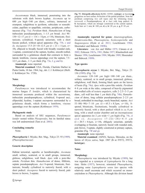
Fig. 72 Ostropella albocincta (K(M): 143941, syntype). a Ascomata<br />
gregarious on host surface. b Section of the partial peridium. Note the<br />
peridium comprising two cell types and the whitening tissue<br />
(arrowed). c Pseudoparaphyses. d, e Asci with long pedicel. f–<br />
h Ascospores, which are strongly constricted at the central septum.<br />
Scale bars: a=1 mm, b=100 μm, d, e, h=20 μm, c, g, f=10 μm<br />
Anamorphs reported for genus: Amarenographium,<br />
Hendersonia-like, Phaeoseptoria, Scolecosporiella and<br />
Stagonospora (Hyde et al. 2011; Leuchtmann 1984;<br />
Shoemaker and Babcock 1989b).<br />
Literature: von Arx and Müller 1975; Câmara et al.<br />
2002; Eriksson 1967a, 1981; Holm 1957; Khashnobish and<br />
Shearer 1996; Leuchtmann 1984; Miyake 1909; Shoemaker<br />
and Babcock 1989b.<br />
Type species<br />
Phaeosphaeria oryzae I. Miyake, Bot. Mag., Tokyo 23:<br />
136 (1909). (Fig. 74)<br />
Ascomata 120–140 μm high×100–140 μm diam.,<br />
solitary, scattered, or in small groups, immersed, globose,<br />
subglobose, wall black, forming black spots on the leaves<br />
of hosts (Fig. 74a). Apex with a pore-like ostiole. Peridium<br />
4–8 μm wide at the sides, composed of heavily pigmented<br />
thin-walled cells of textura angularis, cells 2–2.5×3–5 μm<br />
diam., cell wall less than 1 μm thick (Fig. 74b). Hamathecium<br />
of dense, long cellular pseudoparaphyses 2–2.5 μm<br />
broad, embedded in mucilage, rarely branched, septate. Asci<br />
53–80(−90)×7–10 μm (x ¼ 65:3 8:3mm, n=10), 8-<br />
spored, bitunicate, fissitunicate, broadly cylindrical to<br />
narrowly fusoid, with a short pedicel which is ca. 8μm<br />
long, with a small ocular chamber and an inconspicuous<br />
apical apparatus (to 2 μm wide×1 μm high) (Fig. 74c, d<br />
and e). Ascospores 17– 22(− 28)×4– 5 μ m<br />
(x ¼ 20:5 4:6mm, n=10), obliquely uniseriate, partially<br />
overlapping or biseriate, narrowly fusoid with rounded ends,<br />
pale brown, 3-septate, slightly constricted at primary septum,<br />
granulate (Fig. 74f and g).<br />
Anamorph: none reported.<br />
Material examined: JAPAN, Suruya, Shizuoka, on the<br />
leaves of Oryza sativa, Sept. 1907 (S nr F9572, F9573,<br />
lectotype).<br />
Notes<br />
Morphology<br />
Phaeosphaeria was introduced by Miyake (1909), but<br />
was regarded as a synonym of Leptosphaeria for a long<br />
time. Holm (1957), however, reinstated Phaeosphaeria,<br />
assigning some Leptosphaeria sensu lato species with<br />
relatively small ascomata and which occurred on monocotyledons<br />
to Phaeosphaeria. Although this division based<br />
b


