Fungal Diversity 401, nor did Webster & Lucas in the taxonomic and life-history study (Trans. Brit. Myc. Soc. 42, 332– 342. 1959) of this species. The specimen has most of the features described by Webster & Lucas including the presence of the conidial state Microdiplodia henningsii Staritz. I did not see vertical septa in the ascospores. Webster & Lucas note that vertical septa may be occasionally be lacking. The fungus is otherwise as they describe it although some perithecia collapse and appear cupulate.”—by R.A. Shoemaker. Phylogenetic study None. Concluding remarks The substrate of Chaetoplea sensu Barr (1990b) can be herbaceous stalks, decorticated wood or periderm, or old cotton cloth and string, which may indicate its heterogeneous nature. The ascospores seem very much like Phaeosphaeria which may be an earlier name; more details concerning the ascomatal, peridial and hamathecial structures are needed to make any conclusion. Cilioplea Munk, Dansk botanisk Arkiv 15: 113 (1953). (<strong>Pleosporales</strong>, genera incertae sedis) Generic description Habitat terrestrial, saprobic. Ascomata small- to medium-sized, solitary, scattered or in small groups, immersed, globose or subglobose, papilla covered with short and blackish setae, coriaceous. Peridium thin, comprising small heavily pigmented thick-walled cells of textura angularis. Hamathecium of cellular pseudoparaphyses. Asci 8-spored, bitunicate, fissitunicate, broadly clavate, with a short, furcate pedicel, and small ocular chamber. Ascospores fusoid to narrowly fusoid with narrowly rounded ends, pale brown to reddish brown, multi-transverse septa, usually with one longitudinal septum in some central cells, constricted at the primary septum. Anamorphs reported for genus: none. Literature: Barr 1990b, 1992b; Crivelli 1983; Lumbsch and Huhndorf 2007; Müller 1951; Munk 1953, 1957. Type species Cilioplea coronata (Niessl) Munk, Dansk botanisk Arkiv 15: 113 (1953). (Fig. 23) ≡ Pleospora coronata Niessl, Notiz. Pyr.: 16 (1876). Ascomata 170–290 μm high×200–410 μm diam., solitary, scattered, or in small groups, immersed, globose or subglobose, wall black, papilla raised, 50–80 μm high, with short and blackish setae, coriaceous (Fig. 23a). Peridium 9–15 μm thick laterally, up to 28 μm thick at the apex, thinner at the base, 1-layered, composed of small heavily pigmented thickwalled cells of textura angularis, cells up to 4×2.5 μm diam., cell wall 2–3 μm thick, apex cells smaller and walls thicker (Fig. 23b). Hamathecium of long cellular pseudoparaphyses, 2–3 μm broad. Asci (60-)80–108×10–15 μm (x ¼ 85:3 12:1mm, n=10), 8-spored, bitunicate, fissitunicate, broadly clavate, with a short, thick, furcate pedicel, 5– 15 μm long, and a small ocular chamber (to 3 μm wide× 2 μm high) (Fig. 23c and d). Ascospores 21–27.5×5.5– 7.5 μm (x ¼ 24 6:7mm, n=10), biseriate to uniseriate at base, fusoid to narrowly fusoid with narrowly rounded ends, pale reddish brown, 5–7 transverse septa (mostly 5), usually with one longitudinal septum in some central cells, deeply constricted at the median septum, the part above the primary septum shorter and broader, smooth-walled. Anamorph: none reported. Material examined: GERMANY, Hadiberg. on Reseda lutea Hadiberg, 20 Sept. 1875, Niessl (M 175-89-290, lectotype; M 175-89-291, type). Notes Morphology Cilioplea was introduced by Müller (1951) as a subgenus of Pleospora, and this was followed by Munk (1957), who had earlier proposed it as a separate genus typified by C. coronata based on its hairy papilla, clavate asci as well as its “perfectly paraphysoid” (see Munk 1953). A relatively narrow concept of Pleospora was accepted by Crivelli (1983), and four species was assigned under the separate genus Cilioplea, viz. C. coronata, C. genisticola (Fautrey & Lambotte) Crivelli, C. kansensis (Ellis & Everh.) Crivelli and C. nivalis (Niessl) Crivelli. Subsequently, another six species were added (Barr 1990b, 1992b). Currently, ten species are included under Cilioplea. Phylogenetic study None. Concluding remarks The most striking character of Cilioplea is its setose papilla, which has been shown to have no phylogenetic significance in Lentitheciaceae (Zhang et al. 2009a). Cilioplea was assigned under Lophiostomataceae (Lumbsch and Huhndorf 2007), but there is little morphological similarity with the Lophiostomataceae sensu stricto (Zhang et al. 2009a). Thus its familial placement needs further study. Crivellia Shoemaker & Inderb., in Inderbitzin, Shoemaker, O’Neill, Turgeon & Berbee, Can. J. Bot. 84: 1308 (2006). (Pleosporaceae)
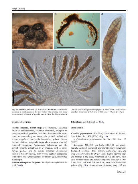
Fungal Diversity Fig. 23 Cilioplea coronata (M 175-89-290, lectotype). a Immersed ascomata in small groups on the host surface (the covering host tissue was removed). b Section of a partial ascoma. Note the thin peridium. c Clavate asci within pseudoparaphyses. d Ascus with a small ocular chamber. Scale bars: a=0.5 mm, b=100 μm, c=50 μm, d=10 μm Generic description Habitat terrestrial, hemibiotrophic or parasitic. Ascomata small- to medium-sized, scattered, immersed, erumpent to nearly superficial, papillate, ostiolate. Peridium thin, composed of two cells types, outer cells of thick walled and textura angularis, inner cells thin-walled, yellow. Hamathecium of dense, long and thin pseudoparaphyses. Asci (4-) 8-spored, bitunicate, fissitunicate dehiscence not observed, broadly cylindrical to cylindrical, with a short, furcate pedicel and an ocular chamber. Ascospores fusoid to broadly fusoid, pale brown, septate, sometimes with one or two vertical septa in the middle cells, constricted at the septa. Anamorphs reported for genus: Brachycladium (Inderbitzin et al. 2006). Literature: Inderbitzin et al. 2006. Type species Crivellia papaveracea (De Not.) Shoemaker & Inderb., Can. J. Bot. 84: 1308 (2006). (Fig. 24) ≡ Cucurbitaria papaveracea De Not., Sfer. Ital.: 62 (1863). Ascomata 210–260 μm high×300–380 μm diam., densely scattered, immersed, erumpent to nearly superficial, flattened globose, dark brown, papillate, ostiolate (Fig. 24a). Peridium 25–30 μm thick, thicker near the apex and thinner at the base, composed of two cell types, outer cells of thick-walled and textura angularis, cells up to 10× 5 μm diam., cell wall 2–4 μm thick, inner cells thin-walled, yellow (Fig. 24b). Hamathecium of dense, long, 1–2 μm
- Page 1 and 2:
Fungal Diversity DOI 10.1007/s13225
- Page 3 and 4:
Fungal Diversity Table 1 Major circ
- Page 5 and 6: Fungal Diversity
- Page 7 and 8: Fungal Diversity biocontrol agent o
- Page 9 and 10: Fungal Diversity substrates and man
- Page 11 and 12: Fungal Diversity 2. To investigate
- Page 13 and 14: Fungal Diversity Table 3 (continued
- Page 15 and 16: Fungal Diversity Table 3 (continued
- Page 17 and 18: Fungal Diversity Table 3 (continued
- Page 19 and 20: Fungal Diversity
- Page 21 and 22: Fungal Diversity Fig. 2 Aigialus gr
- Page 23 and 24: Fungal Diversity Fig. 3 Amniculicol
- Page 25 and 26: Fungal Diversity Literature: Berkel
- Page 27 and 28: Fungal Diversity Ascorhombispora L.
- Page 29 and 30: Fungal Diversity
- Page 31 and 32: Fungal Diversity Fig. 8 Astrosphaer
- Page 33 and 34: Fungal Diversity Fig. 9 Asymmetrico
- Page 35 and 36: Fungal Diversity Notes Morphology B
- Page 37 and 38: Fungal Diversity Generic descriptio
- Page 39 and 40: Fungal Diversity Anamorph: none rep
- Page 41 and 42: Fungal Diversity Fig. 14 Bimuria no
- Page 43 and 44: Fungal Diversity Fig. 15 Bricookea
- Page 45 and 46: Fungal Diversity Fig. 16 Byssolophi
- Page 47 and 48: Fungal Diversity Notes Morphology B
- Page 49 and 50: Fungal Diversity the reaction of pe
- Page 51 and 52: Fungal Diversity
- Page 53 and 54: Fungal Diversity Fig. 21 Chaetomast
- Page 55: Fungal Diversity
- Page 59 and 60: Fungal Diversity with one or two ve
- Page 61 and 62: Fungal Diversity Moreau 1953; Munk
- Page 63 and 64: Fungal Diversity Material examined:
- Page 65 and 66: Fungal Diversity Fig. 28 Dothidotth
- Page 67 and 68: Fungal Diversity Fig. 29 Dubitatio
- Page 69 and 70: Fungal Diversity assigned Entodesmi
- Page 71 and 72: Fungal Diversity fusoid to somewhat
- Page 73 and 74: Fungal Diversity Fig. 33 Hadrospora
- Page 75 and 76: Fungal Diversity Fig. 34 Halotthia
- Page 77 and 78: Fungal Diversity Notes Morphology H
- Page 79 and 80: Fungal Diversity some effused Hypox
- Page 81 and 82: Fungal Diversity Fig. 38 Isthmospor
- Page 83 and 84: Fungal Diversity Fig. 39 Kalmusia e
- Page 85 and 86: Fungal Diversity ascospores were br
- Page 87 and 88: Fungal Diversity furcate pedicel an
- Page 89 and 90: Fungal Diversity Anamorph: none rep
- Page 91 and 92: Fungal Diversity
- Page 93 and 94: Fungal Diversity Material examined:
- Page 95 and 96: Fungal Diversity Fig. 46 Lewia scro
- Page 97 and 98: Fungal Diversity Fig. 47 Lichenopyr
- Page 99 and 100: Fungal Diversity Loculohypoxylon M.
- Page 101 and 102: Fungal Diversity cells small heavil
- Page 103 and 104: Fungal Diversity upper place, septa
- Page 105 and 106: Fungal Diversity
- Page 107 and 108:
Fungal Diversity (CBS 627.86) was i
- Page 109 and 110:
Fungal Diversity Fig. 54 Mamillisph
- Page 111 and 112:
Fungal Diversity Fig. 55 Massarina
- Page 113 and 114:
Fungal Diversity phaeria as a synon
- Page 115 and 116:
Fungal Diversity 5-8 μm diam., ind
- Page 117 and 118:
Fungal Diversity cell wall
- Page 119 and 120:
Fungal Diversity Fig. 60 Mixtura sa
- Page 121 and 122:
Fungal Diversity Fig. 61 Montagnula
- Page 123 and 124:
Fungal Diversity spored, bitunicate
- Page 125 and 126:
Fungal Diversity Fig. 64 Murispora
- Page 127 and 128:
Fungal Diversity Type species Neoph
- Page 129 and 130:
Fungal Diversity brown, 8-septate,
- Page 131 and 132:
Fungal Diversity Fig. 68 Ohleria mo
- Page 133 and 134:
Fungal Diversity Fig. 69 Ohleriella
- Page 135 and 136:
Fungal Diversity Fig. 70 Ophiobolus
- Page 137 and 138:
Fungal Diversity Type species Ostro
- Page 139 and 140:
Fungal Diversity
- Page 141 and 142:
Fungal Diversity (Shoemaker and Bab
- Page 143 and 144:
Fungal Diversity ium thin, composed
- Page 145 and 146:
Fungal Diversity Fig. 76 Platysporo
- Page 147 and 148:
Fungal Diversity Fig. 77 1 Pleomass
- Page 149 and 150:
Fungal Diversity Fig. 78 Pleophragm
- Page 151 and 152:
Fungal Diversity papillate, ostiola
- Page 153 and 154:
Fungal Diversity Williams 1963; Mal
- Page 155 and 156:
Fungal Diversity Generic descriptio
- Page 157 and 158:
Fungal Diversity composed of one ce
- Page 159 and 160:
Fungal Diversity Fig. 84 Saccharico
- Page 161 and 162:
Fungal Diversity and nearly black a
- Page 163 and 164:
Fungal Diversity dense, long trabec
- Page 165 and 166:
Fungal Diversity
- Page 167 and 168:
Fungal Diversity
- Page 169 and 170:
Fungal Diversity Anamorphs reported
- Page 171 and 172:
Fungal Diversity
- Page 173 and 174:
Fungal Diversity
- Page 175 and 176:
Fungal Diversity Fig. 94 Westerdyke
- Page 177 and 178:
Fungal Diversity Fig. 95 Wettsteini
- Page 179 and 180:
Fungal Diversity Fig. 96 Wilmia bra
- Page 181 and 182:
Fungal Diversity Current name: Astr
- Page 183 and 184:
Fungal Diversity spores are actuall
- Page 185 and 186:
Fungal Diversity Fig. 100 Sporormie
- Page 187 and 188:
Fungal Diversity
- Page 189 and 190:
Fungal Diversity Fig. 102 Kriegerie
- Page 191 and 192:
Fungal Diversity Phylogenetic study
- Page 193 and 194:
Fungal Diversity Fig. 104 Zeuctomor
- Page 195 and 196:
Fungal Diversity Fig. 105 Muroia ni
- Page 197 and 198:
Fungal Diversity pseudoparenchymato
- Page 199 and 200:
Fungal Diversity Eremodothis Arx, K
- Page 201 and 202:
Fungal Diversity Type species: Macr
- Page 203 and 204:
Fungal Diversity ascospores of Plat
- Page 205 and 206:
Fungal Diversity monoceras Alcorn n
- Page 207 and 208:
Fungal Diversity tomataceae, Melano
- Page 209 and 210:
Fungal Diversity Table 4 (continued
- Page 211 and 212:
Fungal Diversity 1987b). Based on a
- Page 213 and 214:
Fungal Diversity only do so under v
- Page 215 and 216:
Fungal Diversity Dennis RWG (1968)
- Page 217 and 218:
Fungal Diversity Kirk PM, Cannon PF
- Page 219 and 220:
Fungal Diversity Saccardo PA (1880)
- Page 221:
Fungal Diversity Winter G (1887) As