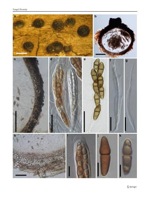
Fungal Diversity Ascospores 16.5–23×7.5–10 μm, biseriate, ovoid or ellipsoidal, dark brown, 1-septate, constricted at the septum, verrucose or verruculose, with or without germ pore (Fig. 93b). Anamorph: none reported. Material examined: SEYCHELLES, Victoria, on submerged branch of Rhizophora mangle L., Mar. 2004, K.D. Hyde (KDH 2137, slide). Notes Morphology Verruculina was introduced to accommodate an obligate marine species, i.e. Verruculina enalia (Kohlmeyer and Volkmann-Kohlmeyer 1990). Verruculina is characterized by immersed, clypeate, carbonaceous, ostiolate and papillate ascomata. The peridium is composed of cells of textura angularis. Pseudoparaphyses are trabeculate and embedded in mucilage. Asci are 8-spored, cylindrical with short pedicels and ocular chamber, and ascospores are ellipsoidal, 1-septate, dark brown, verrucose or verruculose. The partly or completely immersed clypeate ascomata of V. enalia is comparable with those of Didymosphaeria futilis, but it differs from the later by the dark peridium, gelatinous matrix around the pseudoparaphyses, stipitate asci with an ocular chamber, and the verruculose ascospores (Kohlmeyer and Volkmann- Kohlmeyer 1990). Phylogenetic study Based on multigene phylogenetic analysis, Verruculina enalia nested within Testudinaceae (Suetrong et al. 2009). Thus, its familial placement seems clarified. Concluding remarks None. Westerdykella Stolk, Trans. Br. Mycol. Soc. 38: 422 (1955). (Sporormiaceae) Generic description Habitat terrestrial, saprobic (coprophilous). Ascomata small, scattered on the upper layer of the culture medium, wall black. Peridium thin, composed of one layer of cells of polygonal, dark brown, thick-walled cells. Hamathecium not observed. Asci 32-spored, bitunicate nature undetermined, fissitunicate dehiscence not observed, subglobose to ellipsoid, arranged in the centre of the ascomata, with or without a short pedicel. Ascospores globose, brown, 1-celled, without germ pore. Anamorphs reported for genus: Phoma-like (von Arx 1974). Literature: von Arx 1973, 1981; Kruys et al. 2006; Kruys and Wedin 2009; Stolk 1955a. Fig. 92 Trematosphaeria pertusa (a, d, f–i from epitype, b, c, e, j b from neotype). a Ascomata on the host surface. b Section of an ascoma. c, h Section of the peridium. c shows the peridium structure at sides, and h indicates the basal peridium structure. Note the hyaline and thinwalled cells in (h). d Asci amongst pseudoparaphyses. e Ascus with pedicle. f, g Dehiscent ascus. i Upper part of the ascus, showing the ocular chamber and the mucilage covering the apex. j, k Ascospores. Scale bars: a=0.5 mm, b, c=100 μm, d–h=20 μm, i–k=10 μm Type species Westerdykella ornata Stolk, Trans. Br. Mycol. Soc. 38: 422 (1955). (Fig. 94) Ascomata 100–300 μm diam., cleistothecoid, scattered on the upper layer of the culture medium, wall black (Fig. 94a). Peridium composed of one layer of cells of polygonal in front view, dark brown, thick-walled cells, ca. 5μm diam. Hamathecium not observed. Asci 25–32× 16–22 μm, 32-spored, bitunicate nature undetermined, fissitunicate dehiscence not observed, subglobose to ellipsoid, arranged in the centre of the ascomata, with a short furcate pedicel best seen in immature asci (Fig. 94b, c, d and f). Ascospores 6.2–7×6–6.8 μm, globose, brown, 1-celled, ornamented with irregular spiral bands, which occur in four to five coils, without germ pore (Fig. 94e). Anamorph: none reported. On MEA colonies spreading, but somewhat erumpent, with moderate aerial mycelium and even, lobate margins; surface dirty white with luteous to orange patches; reverse orange to sienna. On PDA similar but with sparse aerial mycelium; surface with patches of orange to luteous and dirty white; reverse luteous with cream margins. On OA flat, spreading with sparse aerial mycelium; surface with luteous and dirty white patches and transparent margins; sporulating on OA, visible as black masses of aggregated ascomata; colonies reaching 4 cm diam. on all media (based on <strong>CBS</strong> 379.55). Material examined: MOZAMBIQUE, Inhaca, leg. H.J. Swart, mangrove mud (<strong>CBS</strong> 379.55, holotype). Notes Morphology Westerdykella was introduced to accommodate a coprophilous fungus, which is characterized by cleistothecioid and membraneous ascomata (Stolk 1955a). Asci are subglobose to ellipsoid, stalked, many-spored and evanescent. Ascospores are globose to subglobose, brown, ornamented with spiral bands, without germ pores (Stolk 1955a). Westerdykella was assigned under Phaeosporeae of the Eurotiaceae (Stolk 1955a), and was assigned to Sporormiaceae by von Arx and Müller (1975). Based on the spore ornamentation, von Arx and
Fungal Diversity
- Page 1 and 2:
Fungal Diversity DOI 10.1007/s13225
- Page 3 and 4:
Fungal Diversity Table 1 Major circ
- Page 5 and 6:
Fungal Diversity
- Page 7 and 8:
Fungal Diversity biocontrol agent o
- Page 9 and 10:
Fungal Diversity substrates and man
- Page 11 and 12:
Fungal Diversity 2. To investigate
- Page 13 and 14:
Fungal Diversity Table 3 (continued
- Page 15 and 16:
Fungal Diversity Table 3 (continued
- Page 17 and 18:
Fungal Diversity Table 3 (continued
- Page 19 and 20:
Fungal Diversity
- Page 21 and 22:
Fungal Diversity Fig. 2 Aigialus gr
- Page 23 and 24:
Fungal Diversity Fig. 3 Amniculicol
- Page 25 and 26:
Fungal Diversity Literature: Berkel
- Page 27 and 28:
Fungal Diversity Ascorhombispora L.
- Page 29 and 30:
Fungal Diversity
- Page 31 and 32:
Fungal Diversity Fig. 8 Astrosphaer
- Page 33 and 34:
Fungal Diversity Fig. 9 Asymmetrico
- Page 35 and 36:
Fungal Diversity Notes Morphology B
- Page 37 and 38:
Fungal Diversity Generic descriptio
- Page 39 and 40:
Fungal Diversity Anamorph: none rep
- Page 41 and 42:
Fungal Diversity Fig. 14 Bimuria no
- Page 43 and 44:
Fungal Diversity Fig. 15 Bricookea
- Page 45 and 46:
Fungal Diversity Fig. 16 Byssolophi
- Page 47 and 48:
Fungal Diversity Notes Morphology B
- Page 49 and 50:
Fungal Diversity the reaction of pe
- Page 51 and 52:
Fungal Diversity
- Page 53 and 54:
Fungal Diversity Fig. 21 Chaetomast
- Page 55 and 56:
Fungal Diversity
- Page 57 and 58:
Fungal Diversity Fig. 23 Cilioplea
- Page 59 and 60:
Fungal Diversity with one or two ve
- Page 61 and 62:
Fungal Diversity Moreau 1953; Munk
- Page 63 and 64:
Fungal Diversity Material examined:
- Page 65 and 66:
Fungal Diversity Fig. 28 Dothidotth
- Page 67 and 68:
Fungal Diversity Fig. 29 Dubitatio
- Page 69 and 70:
Fungal Diversity assigned Entodesmi
- Page 71 and 72:
Fungal Diversity fusoid to somewhat
- Page 73 and 74:
Fungal Diversity Fig. 33 Hadrospora
- Page 75 and 76:
Fungal Diversity Fig. 34 Halotthia
- Page 77 and 78:
Fungal Diversity Notes Morphology H
- Page 79 and 80:
Fungal Diversity some effused Hypox
- Page 81 and 82:
Fungal Diversity Fig. 38 Isthmospor
- Page 83 and 84:
Fungal Diversity Fig. 39 Kalmusia e
- Page 85 and 86:
Fungal Diversity ascospores were br
- Page 87 and 88:
Fungal Diversity furcate pedicel an
- Page 89 and 90:
Fungal Diversity Anamorph: none rep
- Page 91 and 92:
Fungal Diversity
- Page 93 and 94:
Fungal Diversity Material examined:
- Page 95 and 96:
Fungal Diversity Fig. 46 Lewia scro
- Page 97 and 98:
Fungal Diversity Fig. 47 Lichenopyr
- Page 99 and 100:
Fungal Diversity Loculohypoxylon M.
- Page 101 and 102:
Fungal Diversity cells small heavil
- Page 103 and 104:
Fungal Diversity upper place, septa
- Page 105 and 106:
Fungal Diversity
- Page 107 and 108:
Fungal Diversity (CBS 627.86) was i
- Page 109 and 110:
Fungal Diversity Fig. 54 Mamillisph
- Page 111 and 112:
Fungal Diversity Fig. 55 Massarina
- Page 113 and 114:
Fungal Diversity phaeria as a synon
- Page 115 and 116:
Fungal Diversity 5-8 μm diam., ind
- Page 117 and 118:
Fungal Diversity cell wall
- Page 119 and 120:
Fungal Diversity Fig. 60 Mixtura sa
- Page 121 and 122: Fungal Diversity Fig. 61 Montagnula
- Page 123 and 124: Fungal Diversity spored, bitunicate
- Page 125 and 126: Fungal Diversity Fig. 64 Murispora
- Page 127 and 128: Fungal Diversity Type species Neoph
- Page 129 and 130: Fungal Diversity brown, 8-septate,
- Page 131 and 132: Fungal Diversity Fig. 68 Ohleria mo
- Page 133 and 134: Fungal Diversity Fig. 69 Ohleriella
- Page 135 and 136: Fungal Diversity Fig. 70 Ophiobolus
- Page 137 and 138: Fungal Diversity Type species Ostro
- Page 139 and 140: Fungal Diversity
- Page 141 and 142: Fungal Diversity (Shoemaker and Bab
- Page 143 and 144: Fungal Diversity ium thin, composed
- Page 145 and 146: Fungal Diversity Fig. 76 Platysporo
- Page 147 and 148: Fungal Diversity Fig. 77 1 Pleomass
- Page 149 and 150: Fungal Diversity Fig. 78 Pleophragm
- Page 151 and 152: Fungal Diversity papillate, ostiola
- Page 153 and 154: Fungal Diversity Williams 1963; Mal
- Page 155 and 156: Fungal Diversity Generic descriptio
- Page 157 and 158: Fungal Diversity composed of one ce
- Page 159 and 160: Fungal Diversity Fig. 84 Saccharico
- Page 161 and 162: Fungal Diversity and nearly black a
- Page 163 and 164: Fungal Diversity dense, long trabec
- Page 165 and 166: Fungal Diversity
- Page 167 and 168: Fungal Diversity
- Page 169 and 170: Fungal Diversity Anamorphs reported
- Page 171: Fungal Diversity
- Page 175 and 176: Fungal Diversity Fig. 94 Westerdyke
- Page 177 and 178: Fungal Diversity Fig. 95 Wettsteini
- Page 179 and 180: Fungal Diversity Fig. 96 Wilmia bra
- Page 181 and 182: Fungal Diversity Current name: Astr
- Page 183 and 184: Fungal Diversity spores are actuall
- Page 185 and 186: Fungal Diversity Fig. 100 Sporormie
- Page 187 and 188: Fungal Diversity
- Page 189 and 190: Fungal Diversity Fig. 102 Kriegerie
- Page 191 and 192: Fungal Diversity Phylogenetic study
- Page 193 and 194: Fungal Diversity Fig. 104 Zeuctomor
- Page 195 and 196: Fungal Diversity Fig. 105 Muroia ni
- Page 197 and 198: Fungal Diversity pseudoparenchymato
- Page 199 and 200: Fungal Diversity Eremodothis Arx, K
- Page 201 and 202: Fungal Diversity Type species: Macr
- Page 203 and 204: Fungal Diversity ascospores of Plat
- Page 205 and 206: Fungal Diversity monoceras Alcorn n
- Page 207 and 208: Fungal Diversity tomataceae, Melano
- Page 209 and 210: Fungal Diversity Table 4 (continued
- Page 211 and 212: Fungal Diversity 1987b). Based on a
- Page 213 and 214: Fungal Diversity only do so under v
- Page 215 and 216: Fungal Diversity Dennis RWG (1968)
- Page 217 and 218: Fungal Diversity Kirk PM, Cannon PF
- Page 219 and 220: Fungal Diversity Saccardo PA (1880)
- Page 221: Fungal Diversity Winter G (1887) As