

Pleosporales - CBS - KNAW
Pleosporales - CBS - KNAW
Pleosporales - CBS - KNAW

Create successful ePaper yourself
Turn your PDF publications into a flip-book with our unique Google optimized e-Paper software.
Fungal Diversity<br />
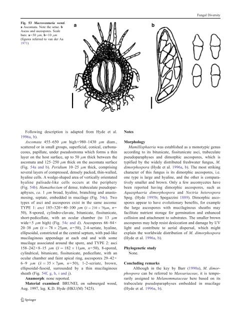
Fig. 53 Macroventuria wenti.<br />
a Ascomata. Note the setae. b<br />
Ascus and ascospores. Scale<br />
bars: a=50 μm, b=10 μm<br />
(figures referred to van der Aa<br />
1971)<br />
Following description is adapted from Hyde et al.<br />
1996a, b).<br />
Ascomata 455–650 μm high×980–1430 μm diam.,<br />
scattered or in small groups, superficial, conical, carbonaceous,<br />
papillate, under pseudostroma which forms a thin<br />
layer on the host surface, up to 50 μm thick between the<br />
ascomata and 125–250 μm thick on the ascomata surface<br />
(Fig. 54a and b). Peridium 10–25 μm thick, comprising<br />
several layers of compressed, densely packed, thin-walled,<br />
hyaline cells. A wedge-shaped area of vertically orientated<br />
hyaline palisade-like cells occurs at the periphery<br />
(Fig. 54b). Hamathecium of dense, trabeculate pseudoparaphyses,<br />
ca. 1μm broad, hyaline, branching and anastomosing,<br />
septate, embedded in mucilage (Fig. 54e). Two<br />
types of asci and ascospores exist in the same ascoma:<br />
TYPE 1: asci 185–320×40–100 μm (x ¼ 210 78mm, n=<br />
50), 8-spored, cylindro-clavate, bitunicate, fissitunicate,<br />
short-pedicellate, with an ocular chamber (to 13 μm<br />
wide×5 μm high) (Fig. 54c and d). Ascospores 66–84×<br />
20–38 μm (x ¼ 78 25mm, n=50), 2-4-seriate, hyaline,<br />
ellipsoidal, constricted at the central septum, with pad-like<br />
mucilaginous appendage at each end and with some<br />
mucilage associated around the spore, and TYPE 2: asci<br />
158–242×8–15 μm (x ¼ 182 11mm, n=50), 8-spored,<br />
cylindrical, bitunicate, fissitunicate, pedicellate, with an<br />
ocular chamber and faint apical ring, ascospores 29–42×<br />
6–9 μm (x ¼ 35 7mm, n =50), 1-2-seriate, brown,<br />
ellipsoidal-fusoid, surrounded by a thin mucilaginous<br />
sheath (Fig. 54f, g, h, i and j).<br />
Anamorph: none reported.<br />
Material examined: BRUNEI, on submerged wood,<br />
Aug. 1997, leg. K.D. Hyde (HKU(M) 7425).<br />
Notes<br />
Morphology<br />
Mamillisphaeria was established as a monotypic genus<br />
according to its bitunicate, fissitunicate asci, trabeculate<br />
pseudoparaphyses and dimorphic ascospores, which is<br />
typified by the widely distributed freshwater fungus, M.<br />
dimorphospora (Hyde et al. 1996a, b). The most striking<br />
character of this fungus is its dimorphic ascospores, i.e.<br />
one type is large and hyaline, and the other is comparatively<br />
smaller and brown. Only a few ascomycetes have<br />
been reported having dimorphic ascospores, such as<br />
Aquasphaeria dimorphospora and Nectria heterospora<br />
Speg. (Hyde 1995b; Spegazzini 1889). Dimorphic ascospores<br />
appear to have evolutionary benefits, for example<br />
the large ascospores with mucilaginous sheaths may<br />
facilitate nutrient storage for germination and enhanced<br />
collision and attachment to substrates. The smaller brown<br />
ascospores may help resist desiccation and damage by UV<br />
light and contribute to aerial dispersal, which might<br />
explain the worldwide distribution of M. dimorphospora<br />
(Hyde et al. 1996a, b).<br />
Phylogenetic study<br />
None.<br />
Concluding remarks<br />
Although in the key by Barr (1990a), M. dimorphospora<br />
can be referred to Massariaceae, itistemporarily<br />
assigned to Melanommataceae here based on its<br />
trabeculate pseudoparaphyses embedded in mucilage<br />
(Hyde et al. 1996a, b).


