

Pleosporales - CBS - KNAW
Pleosporales - CBS - KNAW
Pleosporales - CBS - KNAW

Create successful ePaper yourself
Turn your PDF publications into a flip-book with our unique Google optimized e-Paper software.
Fungal Diversity<br />
papillate, ostiolate. Peridium thin. Hamathecium of dense,<br />
cellular pseudoparaphyses. Asci 8-spored, bitunicate,<br />
fissitunicate, cylindrical to clavate, with furcate pedicel<br />
and small inconspicuous ocular chamber. Ascospores<br />
muriform, brown or pale brown, with or without<br />
sheath.<br />
Anamorphs reported for genus: Stemphylium (Simmons<br />
1985).<br />
Literature: Barr 1981; Frisullo and Braun 1996; Kodsueb<br />
et al. 2006a; Luttrell1951; Wehmeyer1946, 1961, 1975;<br />
Zhang et al. 2009a.<br />
Type species<br />
Pleospora herbarum (Pers.) Rabenh., Klotzschii Herb. Viv.<br />
Mycol. 2: no. 547 (1854). (Fig. 80)<br />
≡ Sphaeria herbarum Pers., Syn. meth. fung. (Göttingen)<br />
1: 78 (1801).<br />
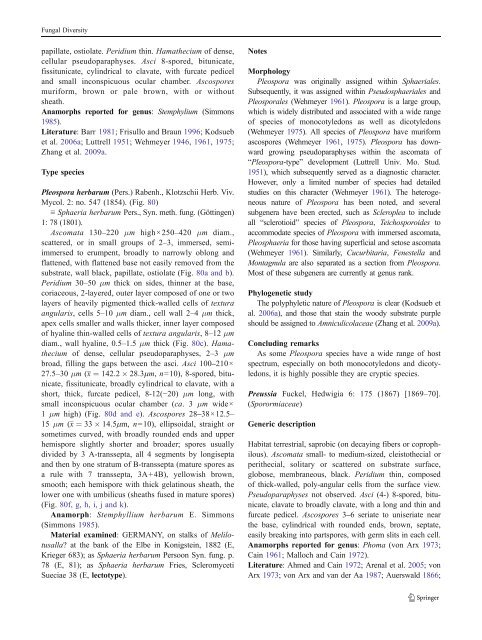
Ascomata 130–220 μm high×250–420 μm diam.,<br />
scattered, or in small groups of 2–3, immersed, semiimmersed<br />
to erumpent, broadly to narrowly oblong and<br />
flattened, with flattened base not easily removed from the<br />
substrate, wall black, papillate, ostiolate (Fig. 80a and b).<br />
Peridium 30–50 μm thick on sides, thinner at the base,<br />
coriaceous, 2-layered, outer layer composed of one or two<br />
layers of heavily pigmented thick-walled cells of textura<br />
angularis, cells 5–10 μm diam., cell wall 2–4 μm thick,<br />
apex cells smaller and walls thicker, inner layer composed<br />
of hyaline thin-walled cells of textura angularis, 8–12 μm<br />
diam., wall hyaline, 0.5–1.5 μm thick (Fig. 80c). Hamathecium<br />
of dense, cellular pseudoparaphyses, 2–3 μm<br />
broad, filling the gaps between the asci. Asci 100–210×<br />
27.5–30 μm (x ¼ 142:2 28:3mm, n=10), 8-spored, bitunicate,<br />
fissitunicate, broadly cylindrical to clavate, with a<br />
short, thick, furcate pedicel, 8-12(−20) μm long, with<br />
small inconspicuous ocular chamber (ca. 3 μm wide×<br />
1 μm high) (Fig. 80d and e). Ascospores 28–38×12.5–<br />
15 μm (x ¼ 33 14:5mm, n=10), ellipsoidal, straight or<br />
sometimes curved, with broadly rounded ends and upper<br />
hemispore slightly shorter and broader; spores usually<br />
divided by 3 A-transsepta, all 4 segments by longisepta<br />
andthenbyonestratumofB-transsepta(maturesporesas<br />
a rule with 7 transsepta, 3A+4B), yellowish brown,<br />
smooth; each hemispore with thick gelatinous sheath, the<br />
lower one with umbilicus (sheaths fused in mature spores)<br />
(Fig. 80f, g, h, i, j and k).<br />
Anamorph: Stemphyllium herbarum E. Simmons<br />
(Simmons 1985).<br />
Material examined: GERMANY, on stalks of Melilotusalla?<br />
at the bank of the Elbe in Konigstein, 1882 (E,<br />
Krieger 683); as Sphaeria herbarum Persoon Syn. fung. p.<br />
78 (E, 81); as Sphaeria herbarum Fries, Scleromyceti<br />
Sueciae 38 (E, lectotype).<br />
Notes<br />
Morphology<br />
Pleospora was originally assigned within Sphaeriales.<br />
Subsequently, it was assigned within Pseudosphaeriales and<br />
<strong>Pleosporales</strong> (Wehmeyer 1961). Pleospora is a large group,<br />
which is widely distributed and associated with a wide range<br />
of species of monocotyledons as well as dicotyledons<br />
(Wehmeyer 1975). All species of Pleospora have muriform<br />
ascospores (Wehmeyer 1961, 1975). Pleospora has downward<br />
growing pseudoparaphyses within the ascomata of<br />
“Pleospora-type” development (Luttrell Univ. Mo. Stud.<br />
1951), which subsequently served as a diagnostic character.<br />
However, only a limited number of species had detailed<br />
studies on this character (Wehmeyer 1961). The heterogeneous<br />
nature of Pleospora has been noted, and several<br />
subgenera have been erected, such as Scleroplea to include<br />
all “sclerotioid” species of Pleospora, Teichosporoides to<br />
accommodate species of Pleospora with immersed ascomata,<br />
Pleosphaeria for those having superficial and setose ascomata<br />
(Wehmeyer 1961). Similarly, Cucurbitaria, Fenestella and<br />
Montagnula are also separated as a section from Pleospora.<br />
Most of these subgenera are currently at genus rank.<br />
Phylogenetic study<br />
The polyphyletic nature of Pleospora is clear (Kodsueb et<br />
al. 2006a), and those that stain the woody substrate purple<br />
should be assigned to Amniculicolaceae (Zhang et al. 2009a).<br />
Concluding remarks<br />
As some Pleospora species have a wide range of host<br />
spectrum, especially on both monocotyledons and dicotyledons,<br />
it is highly possible they are cryptic species.<br />
Preussia Fuckel, Hedwigia 6: 175 (1867) [1869–70].<br />
(Sporormiaceae)<br />
Generic description<br />
Habitat terrestrial, saprobic (on decaying fibers or coprophilous).<br />
Ascomata small- to medium-sized, cleistothecial or<br />
perithecial, solitary or scattered on substrate surface,<br />
globose, membraneous, black. Peridium thin, composed<br />
of thick-walled, poly-angular cells from the surface view.<br />
Pseudoparaphyses not observed. Asci (4-) 8-spored, bitunicate,<br />
clavate to broadly clavate, with a long and thin and<br />
furcate pedicel. Ascospores 3–6 seriate to uniseriate near<br />
the base, cylindrical with rounded ends, brown, septate,<br />
easily breaking into partspores, with germ slits in each cell.<br />
Anamorphs reported for genus: Phoma (von Arx 1973;<br />
Cain 1961; Malloch and Cain 1972).<br />
Literature: Ahmed and Cain 1972; Arenaletal.2005; von<br />
Arx 1973; von Arx and van der Aa 1987; Auerswald1866;


