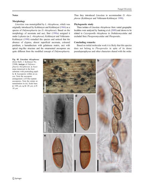
Fungal Diversity Notes Morphology Lineolata was monotypified by L. rhizophorae, whichwas originally introduced by Kohlmeyer and Kohlmeyer (1966)asa species of Didymosphaeria (as D. rhizophorae).Basedonthe morphology of ascomata and asci, Barr (1990a) assignedit under Lojkania (as L. rhizophorae). Kohlmeyer and Volkmann- Kohlmeyer (1990) restudied this species and noticed that the absence of clypeus, almost superficial ascomata, coloured peridium, a hamathecium with gelatinous matrix, asci with apical ring-like structure and the ornamented ascospores are quite different from the modified concept of Didymosphaeria. Thus they introduced Lineolata to accommodate D. rhizophorae (Kohlmeyer and Volkmann-Kohlmeyer 1990). Phylogenetic study Three isolates of Lineolata rhizophorae from varied geographic localities were analyzed by Suetrong et al. (2009)andshowntobe related to Caryosporella rhizophorae in Dothideomycetidae and excluded from Pleosporomycetidae and <strong>Pleosporales</strong>. Concluding remarks Based on initial molecular work it is likely that this species does not belong to <strong>Pleosporales</strong> in spite of its dense pseudoparaphyses and other characters shared with the order. Fig. 48 Lineolata rhizophorae (from Herb. J. Kolmeyer No. 2390b, isotype of Didymosphaeria rhizophorae). a Ascomata immersed in the host substrate with protruding papilla. b Ascospores within an ascus. Note the ascospore arrangement. c–f One-septate ascospores. Note the striate ornamentation in (c). Scale bars: a=100 μm, a, b=20 μm, c–f= 10 μm
Fungal Diversity Loculohypoxylon M.E. Barr, Mycotaxon 3: 326 (1976). (Teichosporaceae) Generic description Habitat terrestrial, saprobic. Ascomata relatively small, gregarious, immersed to erumpent, globose or subglobose, forming under a clypeus, papillate, ostiolate. Peridium thin, a single layer comprising hyaline thinwalled cells of textura angularis or textura prismatica. Hamathecium of septate pseudoparaphyses. Asci (2–4-)8- spored, bitunicate, cylindrical to cylindro-clavate, with a short, furcate pedicel, and wide ocular chamber. Ascospores broadly elliptic to subglobose, often apiculate at both ends, pale to dark brown, aseptate, with a germ slit. Anamorphs reported for genus: none. Literature: von Arx and Müller 1975; Barr 1976. Type species Loculohypoxylon grandineum (Berk. & Rav.) Barr, Mycotaxon 3: 326 (1976). (Fig. 49) ≡ Diatrype grandinea Berk. & Rav., in Berkeley, Grevillea 4: 95 (1876). Ascomata 85–130 μm high×75–145 μm diam., gregarious, immersed to widely erumpent, globose or subglobose, under a reddish brown to black clypeus, papillate, ostiolate (Fig. 49a and b). Peridium 18–30 μm thick laterally, 1-layered, composed of hyaline thin-walled cells of textura angularis to prismatica, cells up to 5×9 μm diam., cell wall 0.5–1 μm thick, apex cells smaller and walls thicker (Fig. 49c). Hamathecium comprising 2–3 μm broad, septate pseudoparaphyses. Asci 70–90×10– 12.5 μm (x ¼ 76:5 10:9mm, n=10), (2–4-)8-spored, bitunicate, cylindrical to cylindro-clavate, with a short, furcate pedicel, up to 25 μm long, with a wide ocular chamber (Fig. 49f, g, and h). Ascospores 7.5–10×5–7 μm (x ¼ 8:3 5:9mm, n=10), uniseriate to partially overlapping at the upper part, broadly elliptic to subglobose, often apiculate at both ends, pale to dark brown, aseptate, with a germ slit (Fig. 49d and e). Anamorph: none reported. Material examined: USA, New Jersey, Newfield, on bark of Quercus coccinea, Sept. 1878, as Diatrype grandinea, Ellis N.A.F. 494 (NY, MASS); on Quercus sp. wood, Nov. 1893, as Anthostoma grandinea B. & Rav., Ellis & Everhart, N.A.F. 494 (NY); Newfield, Oct. 1881, as Diatrype grandinea (NY); Newfield, Jan. 1882, on Quercus coccinea, asDiatrype grandinea B. & Rav, Ex Herb Ellis (NY); Newfield, Nov. 1893, as Anthostoma grandinea, on bark of fallen trunks of Quercus coccinea (NY). Notes Morphology Loculohypoxylon grandineum is one of the rare pleosporalean species having aseptate ascospores. When emphasis is given to ascospore morphology, Semidelitschia (monotypified by S. agasmatica Cain & Luck-Allen) is the most comparable genus. The large ascomata and ascospores, the mucilaginous sheath surrounding the ascospores as well as the coprophilous habitat of S. agasmatica differ from L. grandineum greatly. Thus Loculohypoxylon was introduced as a new genus. Phylogenetic study None. Concluding remarks Aseptate ascospores are rare in <strong>Pleosporales</strong>, and the position of this fungus needs further verification. The familial status of Loculohypoxylon in Teichosporaceae is questionable, as it is simply based on the similarity of living habitat, ascomata and asci with Immotthia and Teichospora (Barr 2002). Lophionema Sacc., Syll. fung. (Abellini) 2: 717 (1883). (<strong>Pleosporales</strong>, genera incertae sedis) Generic description Habitat terrestrial, saprobic? Ascomata solitary, scattered or in small groups, immersed to erumpent, globose to subglobose, with a flattened base, wall black, papillate, ostiolate. Peridium comprising two types of cells which merge in the middle. Hamathecium of trabeculate pseudoparaphyses, septate, rarely anastomosing and branching. Asci 8-spored, bitunicate, fissitunicate unknown, clavate to cylindro-clavate, with a short and furcate pedicel and a small inconspicuous ocular chamber. Ascospores filliform, hyaline to pale yellow, multi-septate, slightly constricted at each septum. Anamorphs reported for genus: none. Literature: Barr1992b; ChestersandBell1970; Ellis and Everhart 1892; Höhnel 1909; Solheim 1949. Type species Lophionema vermisporum (Ellis) Sacc., Syll. fung. (Abellini) 2: 717 (1883). (Fig. 50) ≡ Lophiostoma vermispora Ellis, Bull. Torrey bot. Club 9: 19 (1882). Ascomata 320–430 μm high×280–350 μm diam., solitary, scattered or in small groups of 2–3, immersed to erumpent, globose to subglobose, black, papillate, ostiolate. Papilla 80–120 μm high, up to 150 μm broad, cylindrical to somewhat vertically flattened neck; mostly with a short
- Page 1 and 2:
Fungal Diversity DOI 10.1007/s13225
- Page 3 and 4:
Fungal Diversity Table 1 Major circ
- Page 5 and 6:
Fungal Diversity
- Page 7 and 8:
Fungal Diversity biocontrol agent o
- Page 9 and 10:
Fungal Diversity substrates and man
- Page 11 and 12:
Fungal Diversity 2. To investigate
- Page 13 and 14:
Fungal Diversity Table 3 (continued
- Page 15 and 16:
Fungal Diversity Table 3 (continued
- Page 17 and 18:
Fungal Diversity Table 3 (continued
- Page 19 and 20:
Fungal Diversity
- Page 21 and 22:
Fungal Diversity Fig. 2 Aigialus gr
- Page 23 and 24:
Fungal Diversity Fig. 3 Amniculicol
- Page 25 and 26:
Fungal Diversity Literature: Berkel
- Page 27 and 28:
Fungal Diversity Ascorhombispora L.
- Page 29 and 30:
Fungal Diversity
- Page 31 and 32:
Fungal Diversity Fig. 8 Astrosphaer
- Page 33 and 34:
Fungal Diversity Fig. 9 Asymmetrico
- Page 35 and 36:
Fungal Diversity Notes Morphology B
- Page 37 and 38:
Fungal Diversity Generic descriptio
- Page 39 and 40:
Fungal Diversity Anamorph: none rep
- Page 41 and 42:
Fungal Diversity Fig. 14 Bimuria no
- Page 43 and 44:
Fungal Diversity Fig. 15 Bricookea
- Page 45 and 46:
Fungal Diversity Fig. 16 Byssolophi
- Page 47 and 48: Fungal Diversity Notes Morphology B
- Page 49 and 50: Fungal Diversity the reaction of pe
- Page 51 and 52: Fungal Diversity
- Page 53 and 54: Fungal Diversity Fig. 21 Chaetomast
- Page 55 and 56: Fungal Diversity
- Page 57 and 58: Fungal Diversity Fig. 23 Cilioplea
- Page 59 and 60: Fungal Diversity with one or two ve
- Page 61 and 62: Fungal Diversity Moreau 1953; Munk
- Page 63 and 64: Fungal Diversity Material examined:
- Page 65 and 66: Fungal Diversity Fig. 28 Dothidotth
- Page 67 and 68: Fungal Diversity Fig. 29 Dubitatio
- Page 69 and 70: Fungal Diversity assigned Entodesmi
- Page 71 and 72: Fungal Diversity fusoid to somewhat
- Page 73 and 74: Fungal Diversity Fig. 33 Hadrospora
- Page 75 and 76: Fungal Diversity Fig. 34 Halotthia
- Page 77 and 78: Fungal Diversity Notes Morphology H
- Page 79 and 80: Fungal Diversity some effused Hypox
- Page 81 and 82: Fungal Diversity Fig. 38 Isthmospor
- Page 83 and 84: Fungal Diversity Fig. 39 Kalmusia e
- Page 85 and 86: Fungal Diversity ascospores were br
- Page 87 and 88: Fungal Diversity furcate pedicel an
- Page 89 and 90: Fungal Diversity Anamorph: none rep
- Page 91 and 92: Fungal Diversity
- Page 93 and 94: Fungal Diversity Material examined:
- Page 95 and 96: Fungal Diversity Fig. 46 Lewia scro
- Page 97: Fungal Diversity Fig. 47 Lichenopyr
- Page 101 and 102: Fungal Diversity cells small heavil
- Page 103 and 104: Fungal Diversity upper place, septa
- Page 105 and 106: Fungal Diversity
- Page 107 and 108: Fungal Diversity (CBS 627.86) was i
- Page 109 and 110: Fungal Diversity Fig. 54 Mamillisph
- Page 111 and 112: Fungal Diversity Fig. 55 Massarina
- Page 113 and 114: Fungal Diversity phaeria as a synon
- Page 115 and 116: Fungal Diversity 5-8 μm diam., ind
- Page 117 and 118: Fungal Diversity cell wall
- Page 119 and 120: Fungal Diversity Fig. 60 Mixtura sa
- Page 121 and 122: Fungal Diversity Fig. 61 Montagnula
- Page 123 and 124: Fungal Diversity spored, bitunicate
- Page 125 and 126: Fungal Diversity Fig. 64 Murispora
- Page 127 and 128: Fungal Diversity Type species Neoph
- Page 129 and 130: Fungal Diversity brown, 8-septate,
- Page 131 and 132: Fungal Diversity Fig. 68 Ohleria mo
- Page 133 and 134: Fungal Diversity Fig. 69 Ohleriella
- Page 135 and 136: Fungal Diversity Fig. 70 Ophiobolus
- Page 137 and 138: Fungal Diversity Type species Ostro
- Page 139 and 140: Fungal Diversity
- Page 141 and 142: Fungal Diversity (Shoemaker and Bab
- Page 143 and 144: Fungal Diversity ium thin, composed
- Page 145 and 146: Fungal Diversity Fig. 76 Platysporo
- Page 147 and 148: Fungal Diversity Fig. 77 1 Pleomass
- Page 149 and 150:
Fungal Diversity Fig. 78 Pleophragm
- Page 151 and 152:
Fungal Diversity papillate, ostiola
- Page 153 and 154:
Fungal Diversity Williams 1963; Mal
- Page 155 and 156:
Fungal Diversity Generic descriptio
- Page 157 and 158:
Fungal Diversity composed of one ce
- Page 159 and 160:
Fungal Diversity Fig. 84 Saccharico
- Page 161 and 162:
Fungal Diversity and nearly black a
- Page 163 and 164:
Fungal Diversity dense, long trabec
- Page 165 and 166:
Fungal Diversity
- Page 167 and 168:
Fungal Diversity
- Page 169 and 170:
Fungal Diversity Anamorphs reported
- Page 171 and 172:
Fungal Diversity
- Page 173 and 174:
Fungal Diversity
- Page 175 and 176:
Fungal Diversity Fig. 94 Westerdyke
- Page 177 and 178:
Fungal Diversity Fig. 95 Wettsteini
- Page 179 and 180:
Fungal Diversity Fig. 96 Wilmia bra
- Page 181 and 182:
Fungal Diversity Current name: Astr
- Page 183 and 184:
Fungal Diversity spores are actuall
- Page 185 and 186:
Fungal Diversity Fig. 100 Sporormie
- Page 187 and 188:
Fungal Diversity
- Page 189 and 190:
Fungal Diversity Fig. 102 Kriegerie
- Page 191 and 192:
Fungal Diversity Phylogenetic study
- Page 193 and 194:
Fungal Diversity Fig. 104 Zeuctomor
- Page 195 and 196:
Fungal Diversity Fig. 105 Muroia ni
- Page 197 and 198:
Fungal Diversity pseudoparenchymato
- Page 199 and 200:
Fungal Diversity Eremodothis Arx, K
- Page 201 and 202:
Fungal Diversity Type species: Macr
- Page 203 and 204:
Fungal Diversity ascospores of Plat
- Page 205 and 206:
Fungal Diversity monoceras Alcorn n
- Page 207 and 208:
Fungal Diversity tomataceae, Melano
- Page 209 and 210:
Fungal Diversity Table 4 (continued
- Page 211 and 212:
Fungal Diversity 1987b). Based on a
- Page 213 and 214:
Fungal Diversity only do so under v
- Page 215 and 216:
Fungal Diversity Dennis RWG (1968)
- Page 217 and 218:
Fungal Diversity Kirk PM, Cannon PF
- Page 219 and 220:
Fungal Diversity Saccardo PA (1880)
- Page 221:
Fungal Diversity Winter G (1887) As