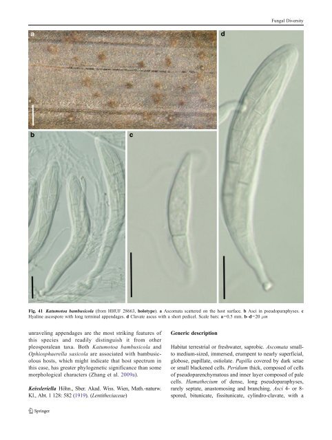
Fungal Diversity Fig. 41 Katumotoa bambusicola (from HHUF 28663, holotype). a Ascomata scattered on the host surface. b Asci in pseudoparaphyses. c Hyaline ascospore with long terminal appendages. d Clavate ascus with a short pedicel. Scale bars: a=0.5 mm. b–d=20 μm unraveling appendages are the most striking features of this species and readily distinguish it from other pleosporalean taxa. Both Katumotoa bambusicola and Ophiosphaerella sasicola are associated with bambusicolous hosts, which might indicate that host spectrum in this case, has greater phylogenetic significance than some morphological characters (Zhang et al. 2009a). Keissleriella Höhn., Sber. Akad. Wiss. Wien, Math.-naturw. Kl., Abt. 1 128: 582 (1919). (Lentitheciaceae) Generic description Habitat terrestrial or freshwater, saprobic. Ascomata smallto medium-sized, immersed, erumpent to nearly superficial, globose, papillate, ostiolate. Papilla covered by dark setae or small blackened cells. Peridium thick, composed of cells of pseudoparenchymatous and inner layer composed of pale cells. Hamathecium of dense, long pseudoparaphyses, rarely septate, anastomosing and branching. Asci 4- or 8- spored, bitunicate, fissitunicate, cylindro-clavate, with a
Fungal Diversity furcate pedicel and a small ocular chamber. Ascospores hyaline to pale brown, ellipsoid to fusoid, 1-septate, constricted at the septum (Barr 1990a). Anamorphs reported for genus: Dendrophoma (Bose 1961). Literature: von Arx and Müller 1975; Bose 1961; Barr 1990a; Dennis 1978; Eriksson 1967a; von Höhnel 1919; Luttrell 1973; Munk 1957; Zhang et al. 2009a. Type species Keissleriella aesculi (Höhn.) Höhn., Sber. Akad. Wiss. Wien, Math.-naturw. Kl., Abt. 1 128: 582 (1919). (Fig. 42) ≡ Pyrenochaeta aesculi Höhn., Ber. dt. bot. Ges. 35: 249 (1917). Ascomata ca. 250μm high×450 μm diam., gregarious, immersed to erumpent, globose or subglobose, with a small black papilla, ca. 75μm high and 110 μm broad, with short black external setae (Fig. 42a). Peridium ca. 25–40 μm wide laterally, up to 70 μm near the apex, thinner at the base, comprising two types of cells which merge in the middle; outer cells composed of small heavily pigmented thick-walled cells, cells ca. 4 μm diam., cell wall up to 4 μm thick, and thick near the apex and thinner laterally and absent in the immersed part of the ascoma, inner cells less pigmented, comprising lightly pigmented to hyaline cells, 5–7 μm thick (Fig. 42a). Hamathecium of dense, long pseudoparaphyses, 0.8–1.2 μm broad, rarely septate, anastomosing and branching, thicker near the base, ca. 2μm, constricted near the septum (Fig. 42b). Asci 80–120×6–11 μm (x ¼ 101 8:5mm, n=10), 4- or 8-spored, bitunicate, fissitunicate, cylindro-clavate, with a furcate pedicel which is up to 20–40 μm long, with a small ocular chamber (Fig. 42e and f). Ascospores 13–18×4–5.5 μm (x ¼ 14:5 4:8mm, n=10), obliquely uniseriate and partially overlapping, fusoid with narrowly rounded ends, hyaline, 1-septate, constricted at the septum, smooth (Fig. 42c and d). Anamorph: none reported. Material examined: AUSTRIA, Brentenmaistal in the Viennese forest, Aesculus hippocastanum L., 1916, Höhnel (FH, holotype of Otthiella aesculi). (Note: only two slides; setae cannot be seen from the slides but could be seen from the drawings on the cover). Notes Morphology Keissleriella is characterized by ascomata with setae in and over the papilla, asci are cylindrical and ascospores are hyaline, 1-septate. Based on the morphological characters, K. aesculi was regarded as conspecific with K. sambucina; as an earlier epithet, K. sambusina typifies the genus (see comments by Barr 1990a). Munk (1957) placed Trichometasphaeria and Keissleriella in Massarinaceae, and distinguished them by their substrates (Trichometasphaeria occurs on herbaceous plants and Keissleriella on woody substrates). Bose (1961) combined Trichometasphaeria under Keissleriella, which was followed by some workers (von Arx and Müller 1975; Dennis 1978; Eriksson 1967a; Luttrell 1973). Barr (1990a), however, maintained these as distinct genera based on the differences of peridium structure and pseudoparaphyses. Phylogenetic study The phylogeny of Keissleriella is poorly studied. Limited phylogenetic information indicates that K. cladophila forms a robust clade with other species of Lentitheciaceae (Zhang et al. 2009a). Concluding remarks The presence of black setae on the surface of papilla is a striking character of Keissleriella, but phylogenetic significance of setae is undetermined yet. Lentithecium K.D. Hyde, J. Fourn. & Yin. Zhang, Fungal Divers. 38: 234 (2009). (Lentitheciaceae) = Tingoldiago K. Hirayama & Kaz. Tanaka, Mycologia 102: 740 (2010) syn. nov. Generic description Habitat freshwater, saprobic. Ascomata small, scattered or gregarious, immersed, slightly erumpent, depressed spherical to lenticular, ostiolate, papillate or epapillate. Peridium thin. Hamathecium of cellular pseudoparaphyses. Asci 8-ascospored, bitunicate, fissitunicate, clavate, short-stipitate. Ascospores broadly fusoid with broadly rounded ends, 1-septate, constricted, hyaline, usually with sheath. Anamorphs reported for genus: none. Literature: Shearer et al. 2009; Zhang et al. 2009a, b. Type species Lentithecium fluviatile (Aptroot & Van Ryck.) K.D. Hyde, J. Fourn. & Yin. Zhang, Fungal Divers. 38: 234 (2009). (Fig. 43) ≡ Massarina fluviatilis Aptroot & Van Ryck., Nova Hedwigia 73: 162 (2001). Ascomata 230–260 μm high×280–325 μm diam., scattered or gregarious, immersed, slightly erumpent, subglobose to depressed spherical, under a small black pseudostroma originating from the apical part of the peridium, apex slightly papillate, ostiole rounded, 60–70 μm diam. (Fig. 43a and b). Peridium 15–20 μm thick at sides and at base, comprising 4–5 layers of angular cells more thick-walled outwards, 50–55 μm
- Page 1 and 2:
Fungal Diversity DOI 10.1007/s13225
- Page 3 and 4:
Fungal Diversity Table 1 Major circ
- Page 5 and 6:
Fungal Diversity
- Page 7 and 8:
Fungal Diversity biocontrol agent o
- Page 9 and 10:
Fungal Diversity substrates and man
- Page 11 and 12:
Fungal Diversity 2. To investigate
- Page 13 and 14:
Fungal Diversity Table 3 (continued
- Page 15 and 16:
Fungal Diversity Table 3 (continued
- Page 17 and 18:
Fungal Diversity Table 3 (continued
- Page 19 and 20:
Fungal Diversity
- Page 21 and 22:
Fungal Diversity Fig. 2 Aigialus gr
- Page 23 and 24:
Fungal Diversity Fig. 3 Amniculicol
- Page 25 and 26:
Fungal Diversity Literature: Berkel
- Page 27 and 28:
Fungal Diversity Ascorhombispora L.
- Page 29 and 30:
Fungal Diversity
- Page 31 and 32:
Fungal Diversity Fig. 8 Astrosphaer
- Page 33 and 34:
Fungal Diversity Fig. 9 Asymmetrico
- Page 35 and 36: Fungal Diversity Notes Morphology B
- Page 37 and 38: Fungal Diversity Generic descriptio
- Page 39 and 40: Fungal Diversity Anamorph: none rep
- Page 41 and 42: Fungal Diversity Fig. 14 Bimuria no
- Page 43 and 44: Fungal Diversity Fig. 15 Bricookea
- Page 45 and 46: Fungal Diversity Fig. 16 Byssolophi
- Page 47 and 48: Fungal Diversity Notes Morphology B
- Page 49 and 50: Fungal Diversity the reaction of pe
- Page 51 and 52: Fungal Diversity
- Page 53 and 54: Fungal Diversity Fig. 21 Chaetomast
- Page 55 and 56: Fungal Diversity
- Page 57 and 58: Fungal Diversity Fig. 23 Cilioplea
- Page 59 and 60: Fungal Diversity with one or two ve
- Page 61 and 62: Fungal Diversity Moreau 1953; Munk
- Page 63 and 64: Fungal Diversity Material examined:
- Page 65 and 66: Fungal Diversity Fig. 28 Dothidotth
- Page 67 and 68: Fungal Diversity Fig. 29 Dubitatio
- Page 69 and 70: Fungal Diversity assigned Entodesmi
- Page 71 and 72: Fungal Diversity fusoid to somewhat
- Page 73 and 74: Fungal Diversity Fig. 33 Hadrospora
- Page 75 and 76: Fungal Diversity Fig. 34 Halotthia
- Page 77 and 78: Fungal Diversity Notes Morphology H
- Page 79 and 80: Fungal Diversity some effused Hypox
- Page 81 and 82: Fungal Diversity Fig. 38 Isthmospor
- Page 83 and 84: Fungal Diversity Fig. 39 Kalmusia e
- Page 85: Fungal Diversity ascospores were br
- Page 89 and 90: Fungal Diversity Anamorph: none rep
- Page 91 and 92: Fungal Diversity
- Page 93 and 94: Fungal Diversity Material examined:
- Page 95 and 96: Fungal Diversity Fig. 46 Lewia scro
- Page 97 and 98: Fungal Diversity Fig. 47 Lichenopyr
- Page 99 and 100: Fungal Diversity Loculohypoxylon M.
- Page 101 and 102: Fungal Diversity cells small heavil
- Page 103 and 104: Fungal Diversity upper place, septa
- Page 105 and 106: Fungal Diversity
- Page 107 and 108: Fungal Diversity (CBS 627.86) was i
- Page 109 and 110: Fungal Diversity Fig. 54 Mamillisph
- Page 111 and 112: Fungal Diversity Fig. 55 Massarina
- Page 113 and 114: Fungal Diversity phaeria as a synon
- Page 115 and 116: Fungal Diversity 5-8 μm diam., ind
- Page 117 and 118: Fungal Diversity cell wall
- Page 119 and 120: Fungal Diversity Fig. 60 Mixtura sa
- Page 121 and 122: Fungal Diversity Fig. 61 Montagnula
- Page 123 and 124: Fungal Diversity spored, bitunicate
- Page 125 and 126: Fungal Diversity Fig. 64 Murispora
- Page 127 and 128: Fungal Diversity Type species Neoph
- Page 129 and 130: Fungal Diversity brown, 8-septate,
- Page 131 and 132: Fungal Diversity Fig. 68 Ohleria mo
- Page 133 and 134: Fungal Diversity Fig. 69 Ohleriella
- Page 135 and 136: Fungal Diversity Fig. 70 Ophiobolus
- Page 137 and 138:
Fungal Diversity Type species Ostro
- Page 139 and 140:
Fungal Diversity
- Page 141 and 142:
Fungal Diversity (Shoemaker and Bab
- Page 143 and 144:
Fungal Diversity ium thin, composed
- Page 145 and 146:
Fungal Diversity Fig. 76 Platysporo
- Page 147 and 148:
Fungal Diversity Fig. 77 1 Pleomass
- Page 149 and 150:
Fungal Diversity Fig. 78 Pleophragm
- Page 151 and 152:
Fungal Diversity papillate, ostiola
- Page 153 and 154:
Fungal Diversity Williams 1963; Mal
- Page 155 and 156:
Fungal Diversity Generic descriptio
- Page 157 and 158:
Fungal Diversity composed of one ce
- Page 159 and 160:
Fungal Diversity Fig. 84 Saccharico
- Page 161 and 162:
Fungal Diversity and nearly black a
- Page 163 and 164:
Fungal Diversity dense, long trabec
- Page 165 and 166:
Fungal Diversity
- Page 167 and 168:
Fungal Diversity
- Page 169 and 170:
Fungal Diversity Anamorphs reported
- Page 171 and 172:
Fungal Diversity
- Page 173 and 174:
Fungal Diversity
- Page 175 and 176:
Fungal Diversity Fig. 94 Westerdyke
- Page 177 and 178:
Fungal Diversity Fig. 95 Wettsteini
- Page 179 and 180:
Fungal Diversity Fig. 96 Wilmia bra
- Page 181 and 182:
Fungal Diversity Current name: Astr
- Page 183 and 184:
Fungal Diversity spores are actuall
- Page 185 and 186:
Fungal Diversity Fig. 100 Sporormie
- Page 187 and 188:
Fungal Diversity
- Page 189 and 190:
Fungal Diversity Fig. 102 Kriegerie
- Page 191 and 192:
Fungal Diversity Phylogenetic study
- Page 193 and 194:
Fungal Diversity Fig. 104 Zeuctomor
- Page 195 and 196:
Fungal Diversity Fig. 105 Muroia ni
- Page 197 and 198:
Fungal Diversity pseudoparenchymato
- Page 199 and 200:
Fungal Diversity Eremodothis Arx, K
- Page 201 and 202:
Fungal Diversity Type species: Macr
- Page 203 and 204:
Fungal Diversity ascospores of Plat
- Page 205 and 206:
Fungal Diversity monoceras Alcorn n
- Page 207 and 208:
Fungal Diversity tomataceae, Melano
- Page 209 and 210:
Fungal Diversity Table 4 (continued
- Page 211 and 212:
Fungal Diversity 1987b). Based on a
- Page 213 and 214:
Fungal Diversity only do so under v
- Page 215 and 216:
Fungal Diversity Dennis RWG (1968)
- Page 217 and 218:
Fungal Diversity Kirk PM, Cannon PF
- Page 219 and 220:
Fungal Diversity Saccardo PA (1880)
- Page 221:
Fungal Diversity Winter G (1887) As