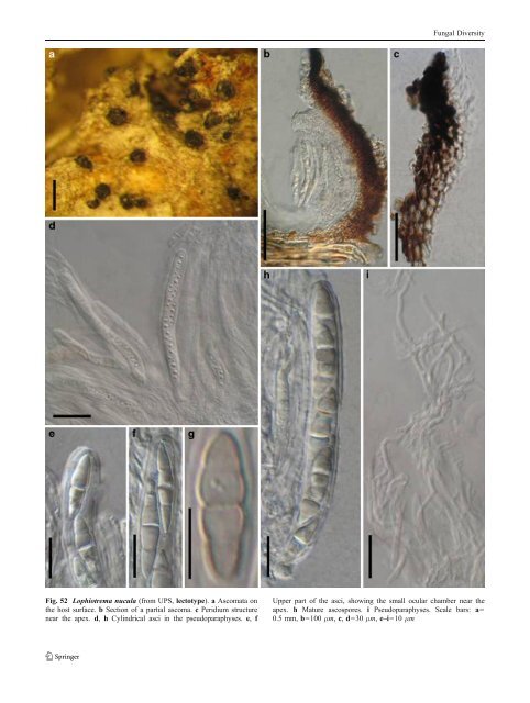
Fungal Diversity Fig. 52 Lophiotrema nucula (from UPS, lectotype). a Ascomata on the host surface. b Section of a partial ascoma. c Peridium structure near the apex. d, h Cylindrical asci in the pseudoparaphyses. e, f Upper part of the asci, showing the small ocular chamber near the apex. h Mature ascospores. i Pseudoparaphyses. Scale bars: a= 0.5 mm, b=100 μm, c, d=30 μm, e–i=10 μm
Fungal Diversity (<strong>CBS</strong> 627.86) was isolated by K. & L. Holm, who had examined the type specimen of L. nucula (Holm and Holm 1988), the culture of Lophiostoma macrostomum used in the analysis are unverified (see comment by Zhang et al. 2009b). For the purpose of this monograph we tentatively maintain Lophiotrema as distinct from Lophiostoma. Macroventuria Aa, Persoonia 6: 359 (1971). (Didymellaceae) Generic description Habitat terrestrial, saprobic. Ascomata small, solitary, scattered, or in groups, initially immersed, becoming erumpent, to nearly superficial, globose to subglobose, roughened with cylindrical setae erect from apex. Peridium thin, membranous. Hamathecium of cellular pseudoparaphyses, seems to easily disappear when mature. Asci bitunicate, somewhat obclavate to fusoid. Ascospores fusoid with broadly to narrowly rounded ends, hyaline, 1-septate, constricted at the septum. Anamorphs reported for genus: none. Literature: van der Aa 1971; von Arx and Müller 1975; Barr 1987a. Type species Macroventuria wentii Aa, Persoonia 6: 361 (1971). (Fig. 53) Ascomata 135–180 μm diam., rarely more than 200 μm diam., solitary, scattered or in groups, initially immersed, becoming erumpent, to nearly superficial, with basal wall remaining immersed in host tissue, globose to subglobose, broadly or narrowly conical, setae erect from the apical region of the ascomata, cylindrical or tapering to the rounded or pointed tip, brown, up to 90 μm long, 5–7.5 μm thick (Fig. 53a). Peridium, 25–35 μm thick, 2-layered, out layer composed of relatively thick-walled cells of textura angularis, cell wall up to 3 μm thick; inner layer cells with a thinner wall and subhyaline; near apex cells smaller (Fig. 53a). Hamathecium of cellular pseudoparaphyses, 1–2 μm thick, evanescing not sure. Asci 75– 93×24–30 μm, 8-spored, without pedicel, bitunicate, somewhat obclavate to fusoid (Fig. 53b). Ascospores 22–32×8–14 μm, 1– 3 seriate, fusoid with broadly to narrowly rounded ends, hyaline, 1-septate, constricted at the septum, smooth (Fig. 53b) (description adapted from van der Aa 1971). Anamorph: none reported. Material referred: USA, Nevada; Death Valley, plant litter, F.W. Went, 229, 1970 (<strong>CBS</strong> 526.71, holotype). Notes Morphology Macroventuria was formally established by van der Aa (1971) represented by M. anomochaeta and M. wentii based on its “near-hyaline, 1-septate ascospores, setose ascomata, and saprobic life style”. Almost all of the above characters (except the saprobic life style) point this group of fungi to Venturiaceae. Thus Macroventuria was assigned to this family as a relatively primitive genus (van der Aa 1971). Subsequently, von Arx and Müller (1975) assigned Macroventuria to Pseudosphaeriaceae (Dothideales), and this proposal was followed by Barr (1987a). Phylogenetic study Phylogenetic analysis based on combined SSU rDNA and LSU rDNA sequences indicated that both of Macroventuria anomochaeta and M. wentii form a robust clade with Leptosphaerulina argentinensis (Speg.) J.H. Graham & Luttr., L. australis, L. trifolii (Rostr.) Petr. and Platychora ulmi, which appear to share phylogenetic affinities with the Leptosphaeriaceae and Phaeosphaeriaceae, but detached from other members of Venturiaceae and Pleosporaceae (Kodsueb et al. 2006a). In addition, culture characters also support the close relationship between Macroventuria and Leptosphaerulina (Barr 1987a). Analysis based on five genes, i.e. SSU, LSU, RPB1, RPB2 andTEF1, indicated Macroventuria anomochaeta resides in the well supported clade of Didymellaceae (Zhang et al. 2009a). Concluding remarks The morphological characters, such as small ascomata and hyaline, 1-septate ascospores all point at Didymellaceae, thus the familial status of Macroventuria is verified. Mamillisphaeria K.D. Hyde, S.W. Wong & E.B.G. Jones, Nova Hedwigia 62: 514 (1996b). (?Melanommataceae) Generic description Habitat freshwater, saprobic. Ascomata superficial, scattered or gregarious, conical, carbonaceous, papillate. Hamathecium of dense, filliform, trabeculate pseudoparaphyses. Asci broadly clavate to clavate, with small ocular chambers and short pedicels. Ascospores of two types, (1): 2-4-seriate, ellipsoid, hyaline, slightly constricted at the main septum; with apical appendages at each end and around the ascospore; (2) 1-2-seriate, ellipsoid to fusoid, brown, with mucilaginous sheath around the ascospore (Hyde et al. 1996b). Anamorphs reported for genus: none. Literature: Hyde et al. 1996a, b. Type species Mamillisphaeria dimorphospora K.D. Hyde, S.W. Wong & E.B.G. Jones, Nova Hedwigia 62: 515 (1996b). (Fig. 54)
- Page 1 and 2:
Fungal Diversity DOI 10.1007/s13225
- Page 3 and 4:
Fungal Diversity Table 1 Major circ
- Page 5 and 6:
Fungal Diversity
- Page 7 and 8:
Fungal Diversity biocontrol agent o
- Page 9 and 10:
Fungal Diversity substrates and man
- Page 11 and 12:
Fungal Diversity 2. To investigate
- Page 13 and 14:
Fungal Diversity Table 3 (continued
- Page 15 and 16:
Fungal Diversity Table 3 (continued
- Page 17 and 18:
Fungal Diversity Table 3 (continued
- Page 19 and 20:
Fungal Diversity
- Page 21 and 22:
Fungal Diversity Fig. 2 Aigialus gr
- Page 23 and 24:
Fungal Diversity Fig. 3 Amniculicol
- Page 25 and 26:
Fungal Diversity Literature: Berkel
- Page 27 and 28:
Fungal Diversity Ascorhombispora L.
- Page 29 and 30:
Fungal Diversity
- Page 31 and 32:
Fungal Diversity Fig. 8 Astrosphaer
- Page 33 and 34:
Fungal Diversity Fig. 9 Asymmetrico
- Page 35 and 36:
Fungal Diversity Notes Morphology B
- Page 37 and 38:
Fungal Diversity Generic descriptio
- Page 39 and 40:
Fungal Diversity Anamorph: none rep
- Page 41 and 42:
Fungal Diversity Fig. 14 Bimuria no
- Page 43 and 44:
Fungal Diversity Fig. 15 Bricookea
- Page 45 and 46:
Fungal Diversity Fig. 16 Byssolophi
- Page 47 and 48:
Fungal Diversity Notes Morphology B
- Page 49 and 50:
Fungal Diversity the reaction of pe
- Page 51 and 52:
Fungal Diversity
- Page 53 and 54:
Fungal Diversity Fig. 21 Chaetomast
- Page 55 and 56: Fungal Diversity
- Page 57 and 58: Fungal Diversity Fig. 23 Cilioplea
- Page 59 and 60: Fungal Diversity with one or two ve
- Page 61 and 62: Fungal Diversity Moreau 1953; Munk
- Page 63 and 64: Fungal Diversity Material examined:
- Page 65 and 66: Fungal Diversity Fig. 28 Dothidotth
- Page 67 and 68: Fungal Diversity Fig. 29 Dubitatio
- Page 69 and 70: Fungal Diversity assigned Entodesmi
- Page 71 and 72: Fungal Diversity fusoid to somewhat
- Page 73 and 74: Fungal Diversity Fig. 33 Hadrospora
- Page 75 and 76: Fungal Diversity Fig. 34 Halotthia
- Page 77 and 78: Fungal Diversity Notes Morphology H
- Page 79 and 80: Fungal Diversity some effused Hypox
- Page 81 and 82: Fungal Diversity Fig. 38 Isthmospor
- Page 83 and 84: Fungal Diversity Fig. 39 Kalmusia e
- Page 85 and 86: Fungal Diversity ascospores were br
- Page 87 and 88: Fungal Diversity furcate pedicel an
- Page 89 and 90: Fungal Diversity Anamorph: none rep
- Page 91 and 92: Fungal Diversity
- Page 93 and 94: Fungal Diversity Material examined:
- Page 95 and 96: Fungal Diversity Fig. 46 Lewia scro
- Page 97 and 98: Fungal Diversity Fig. 47 Lichenopyr
- Page 99 and 100: Fungal Diversity Loculohypoxylon M.
- Page 101 and 102: Fungal Diversity cells small heavil
- Page 103 and 104: Fungal Diversity upper place, septa
- Page 105: Fungal Diversity
- Page 109 and 110: Fungal Diversity Fig. 54 Mamillisph
- Page 111 and 112: Fungal Diversity Fig. 55 Massarina
- Page 113 and 114: Fungal Diversity phaeria as a synon
- Page 115 and 116: Fungal Diversity 5-8 μm diam., ind
- Page 117 and 118: Fungal Diversity cell wall
- Page 119 and 120: Fungal Diversity Fig. 60 Mixtura sa
- Page 121 and 122: Fungal Diversity Fig. 61 Montagnula
- Page 123 and 124: Fungal Diversity spored, bitunicate
- Page 125 and 126: Fungal Diversity Fig. 64 Murispora
- Page 127 and 128: Fungal Diversity Type species Neoph
- Page 129 and 130: Fungal Diversity brown, 8-septate,
- Page 131 and 132: Fungal Diversity Fig. 68 Ohleria mo
- Page 133 and 134: Fungal Diversity Fig. 69 Ohleriella
- Page 135 and 136: Fungal Diversity Fig. 70 Ophiobolus
- Page 137 and 138: Fungal Diversity Type species Ostro
- Page 139 and 140: Fungal Diversity
- Page 141 and 142: Fungal Diversity (Shoemaker and Bab
- Page 143 and 144: Fungal Diversity ium thin, composed
- Page 145 and 146: Fungal Diversity Fig. 76 Platysporo
- Page 147 and 148: Fungal Diversity Fig. 77 1 Pleomass
- Page 149 and 150: Fungal Diversity Fig. 78 Pleophragm
- Page 151 and 152: Fungal Diversity papillate, ostiola
- Page 153 and 154: Fungal Diversity Williams 1963; Mal
- Page 155 and 156: Fungal Diversity Generic descriptio
- Page 157 and 158:
Fungal Diversity composed of one ce
- Page 159 and 160:
Fungal Diversity Fig. 84 Saccharico
- Page 161 and 162:
Fungal Diversity and nearly black a
- Page 163 and 164:
Fungal Diversity dense, long trabec
- Page 165 and 166:
Fungal Diversity
- Page 167 and 168:
Fungal Diversity
- Page 169 and 170:
Fungal Diversity Anamorphs reported
- Page 171 and 172:
Fungal Diversity
- Page 173 and 174:
Fungal Diversity
- Page 175 and 176:
Fungal Diversity Fig. 94 Westerdyke
- Page 177 and 178:
Fungal Diversity Fig. 95 Wettsteini
- Page 179 and 180:
Fungal Diversity Fig. 96 Wilmia bra
- Page 181 and 182:
Fungal Diversity Current name: Astr
- Page 183 and 184:
Fungal Diversity spores are actuall
- Page 185 and 186:
Fungal Diversity Fig. 100 Sporormie
- Page 187 and 188:
Fungal Diversity
- Page 189 and 190:
Fungal Diversity Fig. 102 Kriegerie
- Page 191 and 192:
Fungal Diversity Phylogenetic study
- Page 193 and 194:
Fungal Diversity Fig. 104 Zeuctomor
- Page 195 and 196:
Fungal Diversity Fig. 105 Muroia ni
- Page 197 and 198:
Fungal Diversity pseudoparenchymato
- Page 199 and 200:
Fungal Diversity Eremodothis Arx, K
- Page 201 and 202:
Fungal Diversity Type species: Macr
- Page 203 and 204:
Fungal Diversity ascospores of Plat
- Page 205 and 206:
Fungal Diversity monoceras Alcorn n
- Page 207 and 208:
Fungal Diversity tomataceae, Melano
- Page 209 and 210:
Fungal Diversity Table 4 (continued
- Page 211 and 212:
Fungal Diversity 1987b). Based on a
- Page 213 and 214:
Fungal Diversity only do so under v
- Page 215 and 216:
Fungal Diversity Dennis RWG (1968)
- Page 217 and 218:
Fungal Diversity Kirk PM, Cannon PF
- Page 219 and 220:
Fungal Diversity Saccardo PA (1880)
- Page 221:
Fungal Diversity Winter G (1887) As