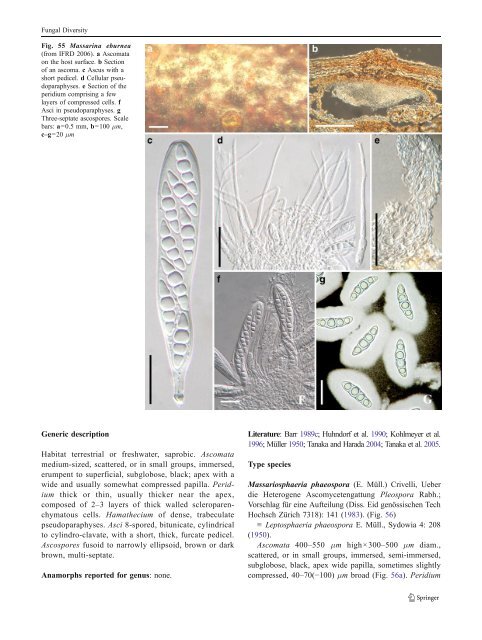
Fungal Diversity Massarina Sacc., Syll. fung. (Abellini) 2: 153 (1883). emend. (Massarinaceae) Generic description Habitat terrestrial, saprobic. Ascomata immersed or superficial, scattered or clustered, globose, conical globose to lenticular, papillate or epapillate, ostiolate. Hamathecium of dense, cellular pseudoparaphyses. Asci clavate to cylindrical, with short pedicels. Ascospores ellipsoid to fusoid, hyaline, 1- to 3-septate, with or without mucilaginous sheath. Anamorphs reported for genus: Ceratophoma (Sivanesan 1984). Literature: Aptroot 1998; Barr 1990a; Bose 1961; Eriksson and Yue 1986; Hyde 1995a; Hyde and Aptroot 1998; Liew et al. 2002; Saccardo 1883; Sivanesan 1984; Tanaka and Harada 2003d; Zhang et al. 2009a, b. Type species Massarina eburnea (Tul. & C. Tul.) Sacc., Syll. fung. (Abellini) 2: 153 (1883). (Fig. 55) ≡ Massaria eburnea Tul. & C. Tul., Sel. Fung. Carp. 2: 239 (1863). Ascomata to 250 μm high×500–700 μm diam., solitary or in small clusters, forming under raised dome-shaped areas, with blackened centres, with a central ostiole, immersed within the cortex of thin dead branches, ellipsoidal, rounded from above, clypeate, neck central, short and barely noticeable on host surface (Fig. 55a). Clypeus ca. 250 μm diam., 60 μm thick, brown, comprising compact brown-walled cells of textura angularis to globulosa beneath host epidermal cells (Fig. 55b). Peridium ca. 20 μm thick comprising 3–5 layers of hyaline compressed cells, fusing at the outside with the host (Fig. 55e). Hamathecium filamentous, cellular pseudoparaphyses, ca. 2 μm broad, septate, embedded in mucilage, without anastomosing (Fig. 55d). Asci 108–170×18–22 μm (x ¼ 144:5 18:8mm, n=10), 8-spored, cylindro-clavate, pedunculate, bitunicate, fissitunicate, (1-)2-seriate, apically rounded, with an ocular chamber and faint ring (J-) (Fig. 55c and f). Ascospores 30–38×8–12 μm (x ¼ 32:4 8:6mm, n= 10), fusoid to ellipsoid, 4-celled, constricted at the septa, hyaline, with acute rounded ends and surrounded by (5– 8 μm diam.) mucilaginous sheath (Fig. 55g). Anamorph: Ceratophoma sp. (Sivanesan 1984). Material examined: FRANCE, on twig of Fagus sp., (Desmazières 1764. P, holotype of Sphaeria pupula var minor), (Mycotheca universalis no. 1951 lectotype). AUS- TRIA, Silesia, Karlsbrunn, on dead twigs of Fagus sylvatica L., Aug. and Sept. 1890, Niessl., De Thümen, sub. Massarina eburnea, ETH. Saxonia, Königsbrunn, on twigs of Fagus sylvatica, Apr. 1882, W. Krieger, Rabenhorst & Winter, Fungi europaei no. 2767, ETH; FRANCE, on a dead twig of Fagus sylvatica, Deux Sèvres, Villiers en Bois, Forêt de Chizé, Rimbaud, 14 Apr. 2008, leg. det. Paul Leroy (IFRD 2006). Notes Morphology Massarina was introduced by Saccardo (1883) for species of pyrenocarpous ascomycetes that had previously been placed in Massaria, but typically had hyaline ascospores (Bose 1961). The family Massarinaceae was described by Munk (1956) to accommodate Massarina. This family was not commonly used and Massarina was later placed within the Lophiostomataceae in the <strong>Pleosporales</strong> (Barr 1990a; Bose 1961; Eriksson and Yue 1986). Of the 160 epithets listed in his monograph, Aptroot accepted only 43 species (Aptroot 1998). The concept of Massarina was widely accepted as having single or aggregated, immersed to erumpent, spherical to hemispherical, pseudothecioid ascomata; cellular pseudoparaphyses; bitunicate, cylindrical to clavate or obpyriform asci; and hyaline, 1–3(−7)-septate, fusoid to long ellipsoid ascospores that mostly have a mucilaginous sheath or appendages (Aptroot 1998; Hyde and Aptroot 1998; Tanaka and Harada 2003d). In the holotype of Sphaeria pupula var. minor (P) and lectotype of Massarina eburnea (ETH), ascospores are reported as “not constricted at the septa” (Hyde 1995a). However, in one of our recent collections, ascospores that are constricted at their septa were observed (Fig. 55g), which was consistent with the description by Fallah and Shearer (2001). This might be because this character is not clear in the old (over 100 years) and dry herbarium specimens, or it may be variable between collections. Phylogenetic study Recent morphological, molecular and anamorphic results indicate, however, that Massarina is polyphyletic (Hyde 1995a; Kirk et al. 2001; Liew et al. 2002). Based on the rDNA dataset, Massarina cisti and the type of Massarina (M. eburnea) forms a robust clade representing Massarina sensu stricto (Zhang et al. 2009a, b). Concluding remarks Massarina sensu stricto should be accepted, which seems to only include some terrestrial and saprobic species. Massariosphaeria (E. Müll.) Crivelli, Diss. Eidgenöss. Techn. Hochschule Zürich 7318: 141 (1983). (?Amniculicolaceae) ≡ Leptosphaeria subgen. Massariosphaeria E. Müll., Sydowia 4: 206 (1950).
Fungal Diversity Fig. 55 Massarina eburnea (from IFRD 2006). a Ascomata on the host surface. b Section of an ascoma. c Ascus with a short pedicel. d Cellular pseudoparaphyses. e Section of the peridium comprising a few layers of compressed cells. f Asci in pseudoparaphyses. g Three-septate ascospores. Scale bars: a=0.5 mm, b=100 μm, c–g=20 μm Generic description Habitat terrestrial or freshwater, saprobic. Ascomata medium-sized, scattered, or in small groups, immersed, erumpent to superficial, subglobose, black; apex with a wide and usually somewhat compressed papilla. Peridium thick or thin, usually thicker near the apex, composed of 2–3 layers of thick walled scleroparenchymatous cells. Hamathecium of dense, trabeculate pseudoparaphyses. Asci 8-spored, bitunicate, cylindrical to cylindro-clavate, with a short, thick, furcate pedicel. Ascospores fusoid to narrowly ellipsoid, brown or dark brown, multi-septate. Anamorphs reported for genus: none. Literature: Barr1989c; Huhndorf et al. 1990; Kohlmeyer et al. 1996; Müller 1950; Tanaka and Harada 2004;Tanakaetal.2005. Type species Massariosphaeria phaeospora (E. Müll.) Crivelli, Ueber die Heterogene Ascomycetengattung Pleospora Rabh.; Vorschlag für eine Aufteilung (Diss. Eid genössischen Tech Hochsch Zürich 7318): 141 (1983). (Fig. 56) ≡ Leptosphaeria phaeospora E. Müll., Sydowia 4: 208 (1950). Ascomata 400–550 μm high×300–500 μm diam., scattered, or in small groups, immersed, semi-immersed, subglobose, black, apex wide papilla, sometimes slightly compressed, 40–70(−100) μm broad (Fig. 56a). Peridium
- Page 1 and 2:
Fungal Diversity DOI 10.1007/s13225
- Page 3 and 4:
Fungal Diversity Table 1 Major circ
- Page 5 and 6:
Fungal Diversity
- Page 7 and 8:
Fungal Diversity biocontrol agent o
- Page 9 and 10:
Fungal Diversity substrates and man
- Page 11 and 12:
Fungal Diversity 2. To investigate
- Page 13 and 14:
Fungal Diversity Table 3 (continued
- Page 15 and 16:
Fungal Diversity Table 3 (continued
- Page 17 and 18:
Fungal Diversity Table 3 (continued
- Page 19 and 20:
Fungal Diversity
- Page 21 and 22:
Fungal Diversity Fig. 2 Aigialus gr
- Page 23 and 24:
Fungal Diversity Fig. 3 Amniculicol
- Page 25 and 26:
Fungal Diversity Literature: Berkel
- Page 27 and 28:
Fungal Diversity Ascorhombispora L.
- Page 29 and 30:
Fungal Diversity
- Page 31 and 32:
Fungal Diversity Fig. 8 Astrosphaer
- Page 33 and 34:
Fungal Diversity Fig. 9 Asymmetrico
- Page 35 and 36:
Fungal Diversity Notes Morphology B
- Page 37 and 38:
Fungal Diversity Generic descriptio
- Page 39 and 40:
Fungal Diversity Anamorph: none rep
- Page 41 and 42:
Fungal Diversity Fig. 14 Bimuria no
- Page 43 and 44:
Fungal Diversity Fig. 15 Bricookea
- Page 45 and 46:
Fungal Diversity Fig. 16 Byssolophi
- Page 47 and 48:
Fungal Diversity Notes Morphology B
- Page 49 and 50:
Fungal Diversity the reaction of pe
- Page 51 and 52:
Fungal Diversity
- Page 53 and 54:
Fungal Diversity Fig. 21 Chaetomast
- Page 55 and 56:
Fungal Diversity
- Page 57 and 58:
Fungal Diversity Fig. 23 Cilioplea
- Page 59 and 60: Fungal Diversity with one or two ve
- Page 61 and 62: Fungal Diversity Moreau 1953; Munk
- Page 63 and 64: Fungal Diversity Material examined:
- Page 65 and 66: Fungal Diversity Fig. 28 Dothidotth
- Page 67 and 68: Fungal Diversity Fig. 29 Dubitatio
- Page 69 and 70: Fungal Diversity assigned Entodesmi
- Page 71 and 72: Fungal Diversity fusoid to somewhat
- Page 73 and 74: Fungal Diversity Fig. 33 Hadrospora
- Page 75 and 76: Fungal Diversity Fig. 34 Halotthia
- Page 77 and 78: Fungal Diversity Notes Morphology H
- Page 79 and 80: Fungal Diversity some effused Hypox
- Page 81 and 82: Fungal Diversity Fig. 38 Isthmospor
- Page 83 and 84: Fungal Diversity Fig. 39 Kalmusia e
- Page 85 and 86: Fungal Diversity ascospores were br
- Page 87 and 88: Fungal Diversity furcate pedicel an
- Page 89 and 90: Fungal Diversity Anamorph: none rep
- Page 91 and 92: Fungal Diversity
- Page 93 and 94: Fungal Diversity Material examined:
- Page 95 and 96: Fungal Diversity Fig. 46 Lewia scro
- Page 97 and 98: Fungal Diversity Fig. 47 Lichenopyr
- Page 99 and 100: Fungal Diversity Loculohypoxylon M.
- Page 101 and 102: Fungal Diversity cells small heavil
- Page 103 and 104: Fungal Diversity upper place, septa
- Page 105 and 106: Fungal Diversity
- Page 107 and 108: Fungal Diversity (CBS 627.86) was i
- Page 109: Fungal Diversity Fig. 54 Mamillisph
- Page 113 and 114: Fungal Diversity phaeria as a synon
- Page 115 and 116: Fungal Diversity 5-8 μm diam., ind
- Page 117 and 118: Fungal Diversity cell wall
- Page 119 and 120: Fungal Diversity Fig. 60 Mixtura sa
- Page 121 and 122: Fungal Diversity Fig. 61 Montagnula
- Page 123 and 124: Fungal Diversity spored, bitunicate
- Page 125 and 126: Fungal Diversity Fig. 64 Murispora
- Page 127 and 128: Fungal Diversity Type species Neoph
- Page 129 and 130: Fungal Diversity brown, 8-septate,
- Page 131 and 132: Fungal Diversity Fig. 68 Ohleria mo
- Page 133 and 134: Fungal Diversity Fig. 69 Ohleriella
- Page 135 and 136: Fungal Diversity Fig. 70 Ophiobolus
- Page 137 and 138: Fungal Diversity Type species Ostro
- Page 139 and 140: Fungal Diversity
- Page 141 and 142: Fungal Diversity (Shoemaker and Bab
- Page 143 and 144: Fungal Diversity ium thin, composed
- Page 145 and 146: Fungal Diversity Fig. 76 Platysporo
- Page 147 and 148: Fungal Diversity Fig. 77 1 Pleomass
- Page 149 and 150: Fungal Diversity Fig. 78 Pleophragm
- Page 151 and 152: Fungal Diversity papillate, ostiola
- Page 153 and 154: Fungal Diversity Williams 1963; Mal
- Page 155 and 156: Fungal Diversity Generic descriptio
- Page 157 and 158: Fungal Diversity composed of one ce
- Page 159 and 160: Fungal Diversity Fig. 84 Saccharico
- Page 161 and 162:
Fungal Diversity and nearly black a
- Page 163 and 164:
Fungal Diversity dense, long trabec
- Page 165 and 166:
Fungal Diversity
- Page 167 and 168:
Fungal Diversity
- Page 169 and 170:
Fungal Diversity Anamorphs reported
- Page 171 and 172:
Fungal Diversity
- Page 173 and 174:
Fungal Diversity
- Page 175 and 176:
Fungal Diversity Fig. 94 Westerdyke
- Page 177 and 178:
Fungal Diversity Fig. 95 Wettsteini
- Page 179 and 180:
Fungal Diversity Fig. 96 Wilmia bra
- Page 181 and 182:
Fungal Diversity Current name: Astr
- Page 183 and 184:
Fungal Diversity spores are actuall
- Page 185 and 186:
Fungal Diversity Fig. 100 Sporormie
- Page 187 and 188:
Fungal Diversity
- Page 189 and 190:
Fungal Diversity Fig. 102 Kriegerie
- Page 191 and 192:
Fungal Diversity Phylogenetic study
- Page 193 and 194:
Fungal Diversity Fig. 104 Zeuctomor
- Page 195 and 196:
Fungal Diversity Fig. 105 Muroia ni
- Page 197 and 198:
Fungal Diversity pseudoparenchymato
- Page 199 and 200:
Fungal Diversity Eremodothis Arx, K
- Page 201 and 202:
Fungal Diversity Type species: Macr
- Page 203 and 204:
Fungal Diversity ascospores of Plat
- Page 205 and 206:
Fungal Diversity monoceras Alcorn n
- Page 207 and 208:
Fungal Diversity tomataceae, Melano
- Page 209 and 210:
Fungal Diversity Table 4 (continued
- Page 211 and 212:
Fungal Diversity 1987b). Based on a
- Page 213 and 214:
Fungal Diversity only do so under v
- Page 215 and 216:
Fungal Diversity Dennis RWG (1968)
- Page 217 and 218:
Fungal Diversity Kirk PM, Cannon PF
- Page 219 and 220:
Fungal Diversity Saccardo PA (1880)
- Page 221:
Fungal Diversity Winter G (1887) As