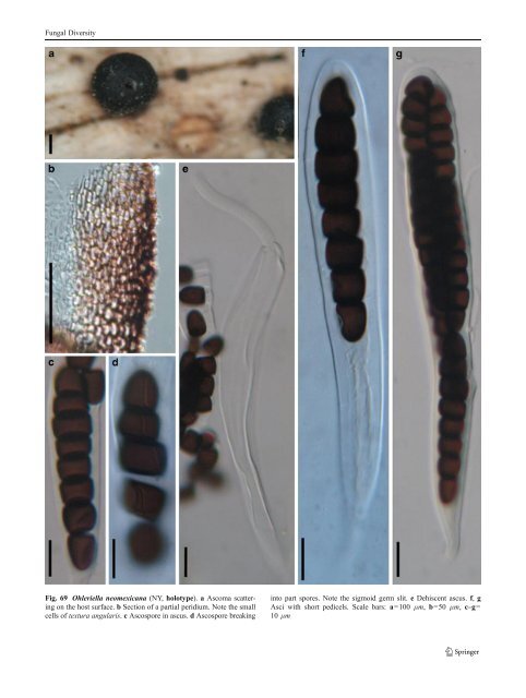
Fungal Diversity immersed in host tissue, coriaceous, globose or subglobose, usually a somewhat thick, short papilla, up to 100 μm high, with a pore-like ostiole (Fig. 69a). Peridium 27–35 μm thick laterally, up to 55 μm thick at the apex, 1- layered, composed of small pigmented cells of textura angularis, cells up to 5×8 μm diam., cell wall 1.5–2 μm thick, apex cells smaller and walls thicker (Fig. 69b). Hamathecium of dense, long trabeculate pseudoparaphyses, 1–1.5 μm broad, anastomosing and branching between and above the asci. Asci 150–208×17.5–25 μm (x ¼ 182:5 22mm, n=10), 8-spored, bitunicate, fissitunicate, cylindrical to cylindro-clavate, with a narrowed, furcate, thin pedicel, 15–55 μm long, 2–3.5 μm broad, with a large truncate ocular chamber best seen in immature asci (to 4 μm wide×3 μm high) (Fig. 69e, f and g). Ascospores 55–72.5× 10⁻¹² μm (x ¼ 63 10:4mm, n=10), 3–4 seriate to uniseriate near the base, cylindrical to clavate, with broadly to narrowly rounded ends, brown, 6–7 transversesepta,easily separating into partspores, with germ slits, central partspores of the ascospores shorter than broad, rectangular in vertical section, round in transverse section, 7–8×6–10 μm diam., apical cells usually longer than broad, 11–17.5×6–7 μm diam. (Fig. 69c and d). Anamorph: none reported. Material examined: USA, Albuquerque, Bernalillo Co., New Mexico, dry gravelly hill, on wood, 29 Nov. 1901, T. S.A. Cockerell (NY, holotype). Notes Morphology Ohleriella was formally established by Earle (1902) based on its “medium to large ascomata with a wide papilla, relatively wide peridium, cylindro-clavate asci, brown to deep brown multi-septate ascospore, with elongated germ slit on each cell”, and was monotypified by O. neomexicana (Barr 1990a). Ohleriella subsequently has been treated as a synonym of Ohleria, Sporormiella or Preussia (Ahmed and Cain 1972; vonArxandMüller1975; Clements and Shear 1931). Spororminula tenerifae, the generic type of Spororminula, was assigned to Ohleriella, thus Spororminula was treated as a synonym of Ohleriella (Barr 1990a). Two new species were introduced by Barr (1990a) from North America. Currently, three species are included in this genus, i.e. O. herculean (Ellis & Everh.) M.E. Barr, O. neomexicana and O. nudilignae M.E. Barr & Malloch (http://www.index fungorum.org; http://www.mycobank.org, 01/03/2009). The generic type, O. neomexicana, is morphologically similar to the coprophilous genus Sporormiella, but is saprobic on grass stems. Phylogenetic study None. Concluding remarks Although we maintain Ohleriella as a separate genus here, its saprobic habitat on grasses and similarity to the coprophilous Sporormiella may indicate a close evolutionary relationship, with the grass saprobic possibly being an early relative of the coprophilous Sporormiella. Alternatively, the species/genera may simply occupy different ecological niches (i.e. dead grass vs dead grass in dung). Molecular studies are needed to resolve this issue. Ophiobolus Reiss, Hedwigia 1:27 (1854). (Phaeosphaeriaceae) Generic description Habitat terrestrial, saprobic or hemibiotrophic. Ascomata medium-sized, solitary, scattered, or in groups, globose or pyriform, coriaceous, black, papillate, ostiolate, periphysate. Peridium thin, thicker near the apex, thinner at the base. Hamathecium of long cellular pseudoparaphyses, septate, anastomosing or branching not observed. Asci 8-spored, bitunicate, fissitunicate dehiscence not observed, cylindrical, with a short, furcate pedicel. Ascospores filamentous, narrower toward the lower end, pale brown, multiseptate, separating into two partspores from the middle septum, from the breaking point, the second cell of each partspore enlarged. Anamorphs reported for genus: Coniothyrium-like, Rhabdospora, Phoma-like and Scolecosporiella (Hyde et al. 2011; Shoemaker 1976; Sivanesan 1984). Literature: Holm 1948, 1957; Müller 1952; Reiss 1854; Shoemaker 1976; Sivanesan 1984. Type species Ophiobolus disseminans Reiss, Hedwigia 1:27 (1854) (Fig. 70). Ascomata 220–380 μm high×290–430 μm diam., solitary, scattered, or in groups often arranged in a row, immersed with a protruding papilla, globose, pyriform, coriaceous, black, periphysate. Papilla 40–90 μm high, with a pore-like ostiole (Fig. 70a and b). Peridium 40–55 μm wide at the sides, up to 70 μm thick at the apex, thinner at the base, comprising two cell types, outer layer composed of small heavily pigmented thick-walled cells of textura angularis, cells 2–5 μm diam., cell wall 2–3 μm thick, apex cells smaller and walls thicker, inner layer composed of lightly pigmented or hyaline thinwalled cells of textura angularis, 5–7 μm diam., wall 1.5– 2 μm thick, merging with pseudoparaphyses (Fig. 70c). Hamathecium of long cellular pseudoparaphyses, 2–3 μm broad, septate, anastomosing or branching not observed (Fig. 70e). Asci 150–195×8–12.5 μm (x ¼ 169:5 10:7mm,
Fungal Diversity Fig. 69 Ohleriella neomexicana (NY, holotype). a Ascoma scattering on the host surface. b Section of a partial peridium. Note the small cells of textura angularis. c Ascospore in ascus. d Ascospore breaking into part spores. Note the sigmoid germ slit. e Dehiscent ascus. f, g Asci with short pedicels. Scale bars: a=100 μm, b=50 μm, c–g= 10 μm
- Page 1 and 2:
Fungal Diversity DOI 10.1007/s13225
- Page 3 and 4:
Fungal Diversity Table 1 Major circ
- Page 5 and 6:
Fungal Diversity
- Page 7 and 8:
Fungal Diversity biocontrol agent o
- Page 9 and 10:
Fungal Diversity substrates and man
- Page 11 and 12:
Fungal Diversity 2. To investigate
- Page 13 and 14:
Fungal Diversity Table 3 (continued
- Page 15 and 16:
Fungal Diversity Table 3 (continued
- Page 17 and 18:
Fungal Diversity Table 3 (continued
- Page 19 and 20:
Fungal Diversity
- Page 21 and 22:
Fungal Diversity Fig. 2 Aigialus gr
- Page 23 and 24:
Fungal Diversity Fig. 3 Amniculicol
- Page 25 and 26:
Fungal Diversity Literature: Berkel
- Page 27 and 28:
Fungal Diversity Ascorhombispora L.
- Page 29 and 30:
Fungal Diversity
- Page 31 and 32:
Fungal Diversity Fig. 8 Astrosphaer
- Page 33 and 34:
Fungal Diversity Fig. 9 Asymmetrico
- Page 35 and 36:
Fungal Diversity Notes Morphology B
- Page 37 and 38:
Fungal Diversity Generic descriptio
- Page 39 and 40:
Fungal Diversity Anamorph: none rep
- Page 41 and 42:
Fungal Diversity Fig. 14 Bimuria no
- Page 43 and 44:
Fungal Diversity Fig. 15 Bricookea
- Page 45 and 46:
Fungal Diversity Fig. 16 Byssolophi
- Page 47 and 48:
Fungal Diversity Notes Morphology B
- Page 49 and 50:
Fungal Diversity the reaction of pe
- Page 51 and 52:
Fungal Diversity
- Page 53 and 54:
Fungal Diversity Fig. 21 Chaetomast
- Page 55 and 56:
Fungal Diversity
- Page 57 and 58:
Fungal Diversity Fig. 23 Cilioplea
- Page 59 and 60:
Fungal Diversity with one or two ve
- Page 61 and 62:
Fungal Diversity Moreau 1953; Munk
- Page 63 and 64:
Fungal Diversity Material examined:
- Page 65 and 66:
Fungal Diversity Fig. 28 Dothidotth
- Page 67 and 68:
Fungal Diversity Fig. 29 Dubitatio
- Page 69 and 70:
Fungal Diversity assigned Entodesmi
- Page 71 and 72:
Fungal Diversity fusoid to somewhat
- Page 73 and 74:
Fungal Diversity Fig. 33 Hadrospora
- Page 75 and 76:
Fungal Diversity Fig. 34 Halotthia
- Page 77 and 78:
Fungal Diversity Notes Morphology H
- Page 79 and 80:
Fungal Diversity some effused Hypox
- Page 81 and 82: Fungal Diversity Fig. 38 Isthmospor
- Page 83 and 84: Fungal Diversity Fig. 39 Kalmusia e
- Page 85 and 86: Fungal Diversity ascospores were br
- Page 87 and 88: Fungal Diversity furcate pedicel an
- Page 89 and 90: Fungal Diversity Anamorph: none rep
- Page 91 and 92: Fungal Diversity
- Page 93 and 94: Fungal Diversity Material examined:
- Page 95 and 96: Fungal Diversity Fig. 46 Lewia scro
- Page 97 and 98: Fungal Diversity Fig. 47 Lichenopyr
- Page 99 and 100: Fungal Diversity Loculohypoxylon M.
- Page 101 and 102: Fungal Diversity cells small heavil
- Page 103 and 104: Fungal Diversity upper place, septa
- Page 105 and 106: Fungal Diversity
- Page 107 and 108: Fungal Diversity (CBS 627.86) was i
- Page 109 and 110: Fungal Diversity Fig. 54 Mamillisph
- Page 111 and 112: Fungal Diversity Fig. 55 Massarina
- Page 113 and 114: Fungal Diversity phaeria as a synon
- Page 115 and 116: Fungal Diversity 5-8 μm diam., ind
- Page 117 and 118: Fungal Diversity cell wall
- Page 119 and 120: Fungal Diversity Fig. 60 Mixtura sa
- Page 121 and 122: Fungal Diversity Fig. 61 Montagnula
- Page 123 and 124: Fungal Diversity spored, bitunicate
- Page 125 and 126: Fungal Diversity Fig. 64 Murispora
- Page 127 and 128: Fungal Diversity Type species Neoph
- Page 129 and 130: Fungal Diversity brown, 8-septate,
- Page 131: Fungal Diversity Fig. 68 Ohleria mo
- Page 135 and 136: Fungal Diversity Fig. 70 Ophiobolus
- Page 137 and 138: Fungal Diversity Type species Ostro
- Page 139 and 140: Fungal Diversity
- Page 141 and 142: Fungal Diversity (Shoemaker and Bab
- Page 143 and 144: Fungal Diversity ium thin, composed
- Page 145 and 146: Fungal Diversity Fig. 76 Platysporo
- Page 147 and 148: Fungal Diversity Fig. 77 1 Pleomass
- Page 149 and 150: Fungal Diversity Fig. 78 Pleophragm
- Page 151 and 152: Fungal Diversity papillate, ostiola
- Page 153 and 154: Fungal Diversity Williams 1963; Mal
- Page 155 and 156: Fungal Diversity Generic descriptio
- Page 157 and 158: Fungal Diversity composed of one ce
- Page 159 and 160: Fungal Diversity Fig. 84 Saccharico
- Page 161 and 162: Fungal Diversity and nearly black a
- Page 163 and 164: Fungal Diversity dense, long trabec
- Page 165 and 166: Fungal Diversity
- Page 167 and 168: Fungal Diversity
- Page 169 and 170: Fungal Diversity Anamorphs reported
- Page 171 and 172: Fungal Diversity
- Page 173 and 174: Fungal Diversity
- Page 175 and 176: Fungal Diversity Fig. 94 Westerdyke
- Page 177 and 178: Fungal Diversity Fig. 95 Wettsteini
- Page 179 and 180: Fungal Diversity Fig. 96 Wilmia bra
- Page 181 and 182: Fungal Diversity Current name: Astr
- Page 183 and 184:
Fungal Diversity spores are actuall
- Page 185 and 186:
Fungal Diversity Fig. 100 Sporormie
- Page 187 and 188:
Fungal Diversity
- Page 189 and 190:
Fungal Diversity Fig. 102 Kriegerie
- Page 191 and 192:
Fungal Diversity Phylogenetic study
- Page 193 and 194:
Fungal Diversity Fig. 104 Zeuctomor
- Page 195 and 196:
Fungal Diversity Fig. 105 Muroia ni
- Page 197 and 198:
Fungal Diversity pseudoparenchymato
- Page 199 and 200:
Fungal Diversity Eremodothis Arx, K
- Page 201 and 202:
Fungal Diversity Type species: Macr
- Page 203 and 204:
Fungal Diversity ascospores of Plat
- Page 205 and 206:
Fungal Diversity monoceras Alcorn n
- Page 207 and 208:
Fungal Diversity tomataceae, Melano
- Page 209 and 210:
Fungal Diversity Table 4 (continued
- Page 211 and 212:
Fungal Diversity 1987b). Based on a
- Page 213 and 214:
Fungal Diversity only do so under v
- Page 215 and 216:
Fungal Diversity Dennis RWG (1968)
- Page 217 and 218:
Fungal Diversity Kirk PM, Cannon PF
- Page 219 and 220:
Fungal Diversity Saccardo PA (1880)
- Page 221:
Fungal Diversity Winter G (1887) As