Fungal Diversity at the septa, dark brown, the apical cells paler with no longitudinal septa, verruculose (Fig. 14e and f). Anamorph: none reported. Material examined: NEW ZEALAND, North Island, Wairarapa District, Nutty Farm, isolated from soil, 3 Mar. 1978, Chea Chark Yen & J.E. Sheridan (<strong>CBS</strong> 107.79, isotype). Notes Morphology Bimuria novae-zelandiae was first isolated from soil of a barley field in New Zealand (Hawksworth et al. 1979). Based on B. novae-zelandiae, the genus is characterized by a very thin peridium, mostly 2-spored and fissitunicate asci as well as the muriform, dark brown, verrucose ascospores (Hawksworth et al. 1979). Because of its unique morphological characters, the familial placement of this genus has been debatable and it has been placed in Pleosporaceae (Hawksworth et al. 1979), in Phaeosphaeriaceae (Barr 1987b)andinMelanommataceae (Lumbsch and Huhndorf 2007). Morphologically, Bimuria is most comparable with some superficially similar or allied genera, in particular Montagnula (Hawksworth et al. 1979). However, the thick carbonaceous peridium distinguishes Montagnula from that of Bimuria (Hawksworth et al. 1979). In addition, the ascospores of Montagnula are discharged forcibly through the ostiole instead of forming a mass outside of the ostiole as in Bimuria (Hawksworth et al. 1979). Ascomauritiana lignicola V.M. Ranghoo & K.D. Hyde has somewhat similar ascospores in 4-spored asci, but this taxon has unitunicate asci (Ranghoo and Hyde 1999). The morphological characters of Bimuria, such as ascospore release and large, thick-walled ascospores may be an adaptation to its soil-borne habitat (Hawksworth et al. 1979). Phylogenetic study Bimuria novae-zelandiae was found to be closely related to Phaeodothis winteri (Niessl) Aptroot (syn. Didymosphaerella opulenta (De Not.) Checa & M.E. Barr) and Montagnula opulenta (De Not.) Aptroot in analysis of combined sequences, i.e. SSU rDNA, LSU rDNA, RPB2 and TEF1 sequences (Schoch et al. 2006, 2009). These two species had been included by Barr (2001) in her new family Montagnulaceae. Concluding remarks We agree with Barr (2001) and include the genus in Montagnulaceae based on both morphological and phylogenetic characters. Bricookea M.E. Barr, Mycotaxon 15: 346 (1982). (?Phaeosphaeriaceae) Generic description Habitat terrestrial, saprobic (or parasitic?). Ascomata small- to medium-sized, solitary, scattered, or in small groups, immersed, erumpent to superficial, depressed globose, papillate, ostiolate. Peridium thin. Hamathecium filliform, cellular pseudoparaphyses, embedded in mucilage, anastomosing, septate. Asci bitunicate, fissitunicate, cylindrical, cylindroclavate or slightly obclavate, with a short knob-like pedicel, with an ocular chamber. Ascospores hyaline, ellipsoid to narrowly obovoid, 3-septate, constricted at each septum. Anamorphs reported for genus: none. Literature:Barr1982a;Berlese1896;Holm1957; Shoemaker and Babcock 1989a. Type species Bricookea sepalorum (Vleugel) M.E. Barr, Mycotaxon 15: 346 (1982). (Fig. 15). ≡ Metasphaeria sepalorum Vleugel, Svensk bot. Tidskr. 2: 369 (1908). Ascomata 120–250 μm high×170–440 μm diam., solitary, scattered, or in small groups, or forming locules in massive stromatic tissues, initially immersed, becoming erumpent, to nearly superficial, depressed globose, black, membraneous, roughened; apex rounded, sometimes very short and almost inconspicuous, with a somewhat slit-like or Y-shaped ostiole (Fig. 15a). Peridium 16–30 μm wide, comprising two types of cells, outer cells heavily pigmented thick-walled textura angularis, cells 4.5–8 μm diam., cell wall 1–1.5 μm thick, inner cells of subhyaline thin-walled textura angularis, cells larger than outer cells (Fig. 15b). Hamathecium of long cellular pseudoparaphyses, 1.5–2 μm broad, embedded in mucilage, anastomosing, septate. Asci 63–83×9.5–11 μm (x ¼ 73:8 10:8mm, n=10), 8-spored, bitunicate, fissitunicate, oblong, cylindro-clavate or slightly obclavate, with a short knob-like pedicel which is 5–13 μm long, with an ocular chamber (Fig. 15c, d and e). Ascospores (14-)15.5–19×5– 7 μm (x ¼ 16:9 5:9mm, n=10), obliquely uniseriate and partially overlapping to biseriate, ellipsoid to narrowly obovoid, hyaline, 3-septate, constricted at each septum, the cells above central septum often broader than the lower ones, smooth (Fig. 15f, g, h, i and j). Anamorph: none reported. Material examined: SWEDEN, on Juncus filliformis, Stockholm, J. Vleugel. Jul. 1907 (S, type as Metasphaeria sepalorum Vleugel). Notes Morphology Bricookea was formally established by Barr (1982a) asa monotypic genus represented by B. sepalorum basedonits
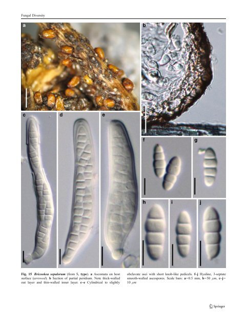
Fungal Diversity Fig. 15 Bricookea sepalorum (from S, type). a Ascomata on host surface (arrowed). b Section of partial peridium. Note thick-walled out layer and thin-walled inner layer. c–e Cylindrical to slightly obclavate asci with short knob-like pedicels. f–j Hyaline, 3-septate smooth-walled ascospores. Scale bars: a=0.5 mm, b=50 μm, c–j= 10 μm
- Page 1 and 2: Fungal Diversity DOI 10.1007/s13225
- Page 3 and 4: Fungal Diversity Table 1 Major circ
- Page 5 and 6: Fungal Diversity
- Page 7 and 8: Fungal Diversity biocontrol agent o
- Page 9 and 10: Fungal Diversity substrates and man
- Page 11 and 12: Fungal Diversity 2. To investigate
- Page 13 and 14: Fungal Diversity Table 3 (continued
- Page 15 and 16: Fungal Diversity Table 3 (continued
- Page 17 and 18: Fungal Diversity Table 3 (continued
- Page 19 and 20: Fungal Diversity
- Page 21 and 22: Fungal Diversity Fig. 2 Aigialus gr
- Page 23 and 24: Fungal Diversity Fig. 3 Amniculicol
- Page 25 and 26: Fungal Diversity Literature: Berkel
- Page 27 and 28: Fungal Diversity Ascorhombispora L.
- Page 29 and 30: Fungal Diversity
- Page 31 and 32: Fungal Diversity Fig. 8 Astrosphaer
- Page 33 and 34: Fungal Diversity Fig. 9 Asymmetrico
- Page 35 and 36: Fungal Diversity Notes Morphology B
- Page 37 and 38: Fungal Diversity Generic descriptio
- Page 39 and 40: Fungal Diversity Anamorph: none rep
- Page 41: Fungal Diversity Fig. 14 Bimuria no
- Page 45 and 46: Fungal Diversity Fig. 16 Byssolophi
- Page 47 and 48: Fungal Diversity Notes Morphology B
- Page 49 and 50: Fungal Diversity the reaction of pe
- Page 51 and 52: Fungal Diversity
- Page 53 and 54: Fungal Diversity Fig. 21 Chaetomast
- Page 55 and 56: Fungal Diversity
- Page 57 and 58: Fungal Diversity Fig. 23 Cilioplea
- Page 59 and 60: Fungal Diversity with one or two ve
- Page 61 and 62: Fungal Diversity Moreau 1953; Munk
- Page 63 and 64: Fungal Diversity Material examined:
- Page 65 and 66: Fungal Diversity Fig. 28 Dothidotth
- Page 67 and 68: Fungal Diversity Fig. 29 Dubitatio
- Page 69 and 70: Fungal Diversity assigned Entodesmi
- Page 71 and 72: Fungal Diversity fusoid to somewhat
- Page 73 and 74: Fungal Diversity Fig. 33 Hadrospora
- Page 75 and 76: Fungal Diversity Fig. 34 Halotthia
- Page 77 and 78: Fungal Diversity Notes Morphology H
- Page 79 and 80: Fungal Diversity some effused Hypox
- Page 81 and 82: Fungal Diversity Fig. 38 Isthmospor
- Page 83 and 84: Fungal Diversity Fig. 39 Kalmusia e
- Page 85 and 86: Fungal Diversity ascospores were br
- Page 87 and 88: Fungal Diversity furcate pedicel an
- Page 89 and 90: Fungal Diversity Anamorph: none rep
- Page 91 and 92: Fungal Diversity
- Page 93 and 94:
Fungal Diversity Material examined:
- Page 95 and 96:
Fungal Diversity Fig. 46 Lewia scro
- Page 97 and 98:
Fungal Diversity Fig. 47 Lichenopyr
- Page 99 and 100:
Fungal Diversity Loculohypoxylon M.
- Page 101 and 102:
Fungal Diversity cells small heavil
- Page 103 and 104:
Fungal Diversity upper place, septa
- Page 105 and 106:
Fungal Diversity
- Page 107 and 108:
Fungal Diversity (CBS 627.86) was i
- Page 109 and 110:
Fungal Diversity Fig. 54 Mamillisph
- Page 111 and 112:
Fungal Diversity Fig. 55 Massarina
- Page 113 and 114:
Fungal Diversity phaeria as a synon
- Page 115 and 116:
Fungal Diversity 5-8 μm diam., ind
- Page 117 and 118:
Fungal Diversity cell wall
- Page 119 and 120:
Fungal Diversity Fig. 60 Mixtura sa
- Page 121 and 122:
Fungal Diversity Fig. 61 Montagnula
- Page 123 and 124:
Fungal Diversity spored, bitunicate
- Page 125 and 126:
Fungal Diversity Fig. 64 Murispora
- Page 127 and 128:
Fungal Diversity Type species Neoph
- Page 129 and 130:
Fungal Diversity brown, 8-septate,
- Page 131 and 132:
Fungal Diversity Fig. 68 Ohleria mo
- Page 133 and 134:
Fungal Diversity Fig. 69 Ohleriella
- Page 135 and 136:
Fungal Diversity Fig. 70 Ophiobolus
- Page 137 and 138:
Fungal Diversity Type species Ostro
- Page 139 and 140:
Fungal Diversity
- Page 141 and 142:
Fungal Diversity (Shoemaker and Bab
- Page 143 and 144:
Fungal Diversity ium thin, composed
- Page 145 and 146:
Fungal Diversity Fig. 76 Platysporo
- Page 147 and 148:
Fungal Diversity Fig. 77 1 Pleomass
- Page 149 and 150:
Fungal Diversity Fig. 78 Pleophragm
- Page 151 and 152:
Fungal Diversity papillate, ostiola
- Page 153 and 154:
Fungal Diversity Williams 1963; Mal
- Page 155 and 156:
Fungal Diversity Generic descriptio
- Page 157 and 158:
Fungal Diversity composed of one ce
- Page 159 and 160:
Fungal Diversity Fig. 84 Saccharico
- Page 161 and 162:
Fungal Diversity and nearly black a
- Page 163 and 164:
Fungal Diversity dense, long trabec
- Page 165 and 166:
Fungal Diversity
- Page 167 and 168:
Fungal Diversity
- Page 169 and 170:
Fungal Diversity Anamorphs reported
- Page 171 and 172:
Fungal Diversity
- Page 173 and 174:
Fungal Diversity
- Page 175 and 176:
Fungal Diversity Fig. 94 Westerdyke
- Page 177 and 178:
Fungal Diversity Fig. 95 Wettsteini
- Page 179 and 180:
Fungal Diversity Fig. 96 Wilmia bra
- Page 181 and 182:
Fungal Diversity Current name: Astr
- Page 183 and 184:
Fungal Diversity spores are actuall
- Page 185 and 186:
Fungal Diversity Fig. 100 Sporormie
- Page 187 and 188:
Fungal Diversity
- Page 189 and 190:
Fungal Diversity Fig. 102 Kriegerie
- Page 191 and 192:
Fungal Diversity Phylogenetic study
- Page 193 and 194:
Fungal Diversity Fig. 104 Zeuctomor
- Page 195 and 196:
Fungal Diversity Fig. 105 Muroia ni
- Page 197 and 198:
Fungal Diversity pseudoparenchymato
- Page 199 and 200:
Fungal Diversity Eremodothis Arx, K
- Page 201 and 202:
Fungal Diversity Type species: Macr
- Page 203 and 204:
Fungal Diversity ascospores of Plat
- Page 205 and 206:
Fungal Diversity monoceras Alcorn n
- Page 207 and 208:
Fungal Diversity tomataceae, Melano
- Page 209 and 210:
Fungal Diversity Table 4 (continued
- Page 211 and 212:
Fungal Diversity 1987b). Based on a
- Page 213 and 214:
Fungal Diversity only do so under v
- Page 215 and 216:
Fungal Diversity Dennis RWG (1968)
- Page 217 and 218:
Fungal Diversity Kirk PM, Cannon PF
- Page 219 and 220:
Fungal Diversity Saccardo PA (1880)
- Page 221:
Fungal Diversity Winter G (1887) As