

Pleosporales - CBS - KNAW
Pleosporales - CBS - KNAW
Pleosporales - CBS - KNAW

Create successful ePaper yourself
Turn your PDF publications into a flip-book with our unique Google optimized e-Paper software.
Fungal Diversity<br />
papillate, ostiolate, periphysate, carbonaceous (Fig. 20a<br />
and b). Peridium 120–150 μm thick at sides, up to<br />
200 μm thick at the apex, thinner at the base, 3-layered,<br />
outer layer composed of golden-yellow, very thickwalled<br />
cells of textura epidermoidea, mixed with subglobose,<br />
large cells near the surface, cells 7–15 μm<br />
diam., middle layer composed of deep brown, very thickwalled<br />
cells of textura epidermoidea, inner layer composed<br />
of hyaline, thin-walled cells of textura prismatica,<br />
up to 50×5 μm diam., merging with pseudoparaphyses<br />
(Fig. 20b, c and d). Hamathecium of dense, long<br />
trabeculate pseudoparaphyses, 1.5-2 μm wide, anastomosing<br />
and branching above the asci. Asci 225–250<br />
(−275)×14–17 μm (x ¼ 137 16:3mm, n=10), 8-spored,<br />
bitunicate, fissitunicate, cylindrical, with a long, narrowed,<br />
pedicel which is up to 75 μm long, apical<br />
characters not observed (Fig. 20e). Ascospores 25–28<br />
(−30)×9–13 μm (x ¼ 26:8 11mm, n=10), uniseriate to<br />
partially overlapping, ellipsoidal to broadly fusoid with<br />
narrow hyaline rounded ends, deep reddish brown, thickwalled,<br />
1-septate with hyaline germ pore at each end, slightly<br />
constricted at the septum, verruculose, sometimes with “net”-<br />
like ridged ornamentations (Fig. 20f and g).<br />
Anamorph: suspected spermatia (Kohlmeyer 1985).<br />
Material examined: BELIZE, Twin Cays, tip of prop<br />
root of Rhizophora mangle, 18 Mar. 1984, J. Kohlmeyer<br />
(NY. Herb. J. Kohlmeyer No. 4532a, holotype).<br />
Notes<br />
Morphology<br />
Caryosporella was formally established by Kohlmeyer<br />
(1985) based on the obligate marine fungus, C. rhizophorae,<br />
which is characterized by its superficial ascomata,<br />
3-layered peridium, filliform trabeculate pseudoparaphyses,<br />
and brown, 1-septate ascospores. Caryosporella was<br />
originally assigned to Massariaceae despite several major<br />
differences, such as the superficial ascomata, reddish<br />
brown ascospores (Kohlmeyer 1985). Subsequently,<br />
Caryosporella was assigned to Melanommataceae<br />
(Eriksson 2006; Lumbsch and Huhndorf 2007).<br />
Phylogenetic study<br />
Suetrong et al. (2009) showed that a single isolate of<br />
Caryosporella rhizophorae does not reside in <strong>Pleosporales</strong>,<br />
but is related to Lineolata rhizophorae (Kohlm. & E.<br />
Kohlm.) Kohlm. & Volkm.-Kohlm. and placed in Dothideomycetidae<br />
incertae sedis.<br />
Concluding remarks<br />
As an obligate marine fungus, the familial placement of<br />
Caryosporella rhizophorae is uncertain but it may not<br />
belong to <strong>Pleosporales</strong>.<br />
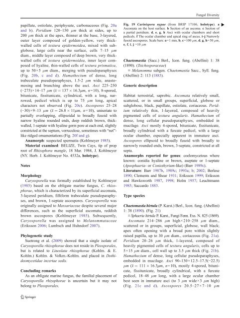
Fig. 19 Carinispora nypae (from BRIP 17106, holotype). a b<br />
Ascomata on the host surface. b Section of an ascoma. c Section of<br />
a partial peridium. d, e, g, h Asci with ocular chambers and short<br />
pedicels. f The ocular chamber and apical ring of ascus. i–j Narrowly<br />
fusoid ascospores. Scale bars: a=1 mm, b, c=100 μm, d, g, h=50 μm,<br />
e, f, i, j =10 μm<br />
Chaetomastia (Sacc.) Berl., Icon. fung. (Abellini) 1: 38<br />
(1890). (Teichosporaceae)<br />
≡ Melanomma subgen. Chaetomastia Sacc., Syll. fung.<br />
(Abellini) 2: 113 (1883).<br />
Generic description<br />
Habitat terrestrial, saprobic. Ascomata relatively small,<br />
scattered, or in small groups, superficial, globose or<br />
subglobose, black, papillate, ostiolate, coriaceous. Peridium<br />
relatively thin, 1-layered, composed of heavily<br />
pigmented cells of textura angularis. Hamathecium of<br />
dense, long cellular pseudoparaphyses, embedded in<br />
mucilage. Asci mostly 4-spored, bitunicate, fissitunicate,<br />
broadly cylindrical with a furcate pedicel, with a large<br />
ocular chamber, especially apparent in immature asci.<br />
Ascospores ellipsoid to broadly fusoid with broadly to<br />
narrowly rounded ends, brown, 3-septate, constricted at all<br />
septa.<br />
Anamorphs reported for genus: coelomycetous where<br />
known: conidia hyaline or brown, aseptate or 1-septate<br />
(Aposphaeria- orConiothyrium-like) (Barr 1989c).<br />
Literature: Barr 1987b, 1989c; 1993a; b; 2002; Berlese<br />
1890; Clements and Shear 1931; Eriksson 1999; Eriksson<br />
and Hawksworth 1987, 1998; Holm 1957; Leuchtmann<br />
1985; Saccardo 1883.<br />
Type species<br />
Chaetomastia hirtula (P. Karst.) Berl., Icon. fung. (Abellini)<br />
1: 38 (1890). (Fig. 21)<br />
≡ Sphaeria hirtula P. Karst., Fungi Fenn. Exs. N. 825 (1869).<br />
Ascomata 214–286 μm high×210–258 μm diam.,<br />
scattered or in groups, superficial, globose, wall black;<br />
apex often opening with a broad pore within slightly<br />
raised papilla, up to 30 μm diam., coriaceous (Fig. 21a).<br />
Peridium 20–26 μm thick, 1-layered, composed of<br />
heavily pigmented cells of textura angularis, cells up to<br />
5×15 μm diam., cell wall up to 3.5 μm thick (Fig. 21b).<br />
Hamathecium of dense, long cellular pseudoparaphyses,<br />
embedded in mucilage. Asci 90–130×12.5–17.5(−22.5)<br />
μm (x ¼ 111 16:3mm, n=10), mostly 4-spored, bitunicate,<br />
fissitunicate, broadly cylindrical, with a furcate<br />
pedicel, 18–48 μm long, with a large ocular chamber<br />
best seen in immature asci (to 3 μm wide×3 μm high)<br />
(Fig. 21c and d). Ascospores 20.5–27×7–10 μm


