

Pleosporales - CBS - KNAW
Pleosporales - CBS - KNAW
Pleosporales - CBS - KNAW

Create successful ePaper yourself
Turn your PDF publications into a flip-book with our unique Google optimized e-Paper software.
Fungal Diversity<br />
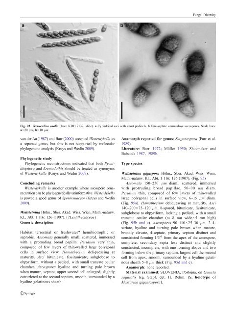
Fig. 93 Verruculina enalia (from KDH 2137, slide). a Cylindrical asci with short pedicels. b One-septate verruculose ascospores. Scale bars:<br />
a=20 μm, b=10 μm<br />
van der Aa (1987) andBarr(2000) acceptedWesterdykella as<br />
a separate genus, but this is not supported by molecular<br />
phylogenetic analysis (Kruys and Wedin 2009).<br />
Phylogenetic study<br />
Phylogenetic reconstructions indicated that both Pycnidiophora<br />
and Eremodothis should be treated as synonyms<br />
of Westerdykella (Kruys and Wedin 2009).<br />
Concluding remarks<br />
Westerdykella is another example where ascospore ornamentation<br />
can be phylogenetically uninformative. Westerdykella<br />
is proved a good genus of Sporormiaceae (Kruys and Wedin<br />
2009).<br />
Wettsteinina Höhn., Sber. Akad. Wiss. Wien, Math.-naturw.<br />
Kl., Abt. I 116: 126 (1907). (?Lentitheciaceae)<br />
Generic description<br />
Habitat terrestrial or freshwater? hemibiotrophic or<br />
saprobic. Ascomata generally small, scattered, immersed<br />
with a protruding broad papilla. Peridium very thin,<br />
composed of few layers of thin-walled large polygonal<br />
cells in surface view. Hamathecium deliquescing at<br />
maturity. Asci bitunicate, fissitunicate, subglobose to<br />
obpyriform, without a pedicel, with small truncate ocular<br />
chamber. Ascospores hyaline and turning pale brown<br />
when mature, septate, upper second cell enlarged, slightly<br />
constricted at the second septum, smooth, surrounded by a<br />
hyaline gelatinous sheath.<br />
Anamorph reported for genus: Stagonospora (Farr et al.<br />
1989).<br />
Literature: Barr 1972; Müller 1950; Shoemaker and<br />
Babcock 1987, 1989b.<br />
Type species<br />
Wettsteinina gigaspora Höhn., Sber. Akad. Wiss. Wien,<br />
Math.-naturw. Kl., Abt. 1 116: 126 (1907). (Fig. 95)<br />
Ascomata 150–250 μm diam., scattered, immersed<br />
with protruding broad papillae, 50–90 μm diam.<br />
Peridium thin, composed of few layers of thin-walled<br />
large polygonal cells in surface view, 6–15 μm diam.<br />
(Fig. 95a). Hamathecium deliquescing at maturity. Asci<br />
140–200×75–120 μm, 8-spored, bitunicate, fissitunicate,<br />
subglobose to obpyriform, lacking a pedicel, with a small<br />
truncate ocular chamber (to 8 μm wide×5 μm high)<br />
(Fig. 95b and c). Ascospores 90–110×25–30 μm, 2–4-<br />
seriate, hyaline and turning pale brown when mature,<br />
broadly clavate, 4-septate, primary septum distinct and<br />
constricted forming 1/3 rd from the apex of the ascospore,<br />
complete, secondary septa less distinct and slightly<br />
constricted, incomplete, with one forming above and two<br />
forming below the primary septum, largest cell the second<br />
cell from apex, smooth, surrounded by a hyaline gelatinous<br />
sheath 5–8 μm thick (Fig. 95d and e).<br />
Anamorph: none reported.<br />
Material examined: SLOVENIA, Postojna, on Genista<br />
sagittalis leg. Stapf. det. H. Rehm. (S, holotype of<br />
Massarina gigantospora).


