

Pleosporales - CBS - KNAW
Pleosporales - CBS - KNAW
Pleosporales - CBS - KNAW

Create successful ePaper yourself
Turn your PDF publications into a flip-book with our unique Google optimized e-Paper software.
Fungal Diversity<br />
5–8 μm diam., individual cell wall to 1.5–2 μm thick, in<br />
places with columns of textura prismatica, and larger, paler<br />
cells of textura prismatica towards the interior and at the<br />
base (Fig. 58b). Hamathecium of dense, filamentous, 1–2<br />
(−2.5) μm broad, branching, rarely anastomosing, septate<br />
pseudoparaphyses. Asci 98–123×6.5–7.5(−9) μm<br />
(x ¼ 109 7:5mm, n=10), 8-spored, bitunicate, fissitunicate,<br />
cylindrical to fusoid, with a short, furcate pedicel, to 25 μm<br />
long, with an ocular chamber (Fig. 58c, d, e, f and g).<br />
Ascospores 14–17.5(−19)×4.5–6.5 μm (x ¼ 15:8 5:2mm,<br />
n=10), obliquely uniseriate and partially overlapping, broadly<br />
fusoid to fusoid with broadly rounded ends, straight or slightly<br />
curved, smooth, olive-brown, 4-celled, slightly constricted at<br />
the septa, the second cell from the top slightly wider than the<br />
others, no sheath (Fig. 58h,i,j,kandl).<br />
Anamorph: Aposphaeria agminalis Sacc. or Phoma<br />
agminalis Sacc. (Sivanesan 1984).<br />
Colonies (of epitype) reaching 4 cm diam. after 20 days<br />
growth on PDA at 25°C, depressed to raised, cottony to woolly,<br />
with rhizoidal margin, grey, reverse darkened. Phoma-like<br />
anamorph has been reported by Chesters (1938) and<br />
Sivanesan (1984), but no anamorphic stage was observed<br />
in the cultures of IFRDCC 2044, <strong>CBS</strong> 109.77 and <strong>CBS</strong><br />
371.75 after culturing 3 months on PDA.<br />
Material examined:ondecayingwood(UPS,Scler.suec.<br />
n. 120, holotype,asSphaeria pulvis-pyrius Pers.); FRANCE,<br />
Ariège, Rimont, Saurine, on bark of Salix caprea, 10Apr.<br />
2008, Jacques Fournier (IFRD 2001, epitype).<br />
Notes<br />
Morphology<br />
Melanomma, the familial type of Melanommataceae, was<br />
formally established by Fuckel (1870, p 159) based on its small,<br />
carbonaceous ascomata, having: “sporen meist 2–3 mal septirt,<br />
selten ohne Scheidewand, braun oder wasscrhell.” (Chesters<br />
1938; Fuckel 1870). Saccardo (1878, p. 344) emended this<br />
genus as “Spores ovate or oblong, multi-septate, coloured.”<br />
Subsequently, Saccardo (1883, p. 98) extended the description<br />
of Melanomma as “Perithecia gregarious, seldom scattered,<br />
somewhat superficial, sphaerical, papillate or blunt, carbonaceous,<br />
smooth or somewhat hairy. Asci elongate, for the most<br />
part accompanied by paraphyses, 8-spored. Spores oblong or<br />
somewhat spindle-shaped, two to many septate, olive or dark<br />
brown. Species of Sphaeria belong here for the most part.”<br />
Melanomma pulvis-pyrius was erected as the lectotype<br />
species (Barr 1990a; Chesters1938). Barr (1990a) gavea<br />
detailed circumscription for Melanomma, under which<br />
Melanomma contains about 20 species (Kirk et al. 2001).<br />
Melanomma pulvis-pyrius is characterized by its gregarious,<br />
superficial ascomata with short papillate, cylindrical asci with a<br />
short pedicel and fusoid, olive-brown, 3-septate ascospores<br />
(Chesters 1938; Zhang et al. 2008a). One of the diagnostic<br />
characters of Melanommataceae is the trabeculate pseudoparaphyses,<br />
although no typical trabeculate pseudoparaphyses could<br />
be found in the neotype (Scler. suec. n. 120, UPS) and epitype<br />
(IFRD 2001) of M. pulvis-pyrius (Zhang et al. 2008a).<br />
Phylogenetic study<br />
Phylogenetic analysis based on five genes (LSU, SSU,<br />
RPB1, RPB2 andEF1) indicates that Melanomma pulvispyrius<br />
forms a robust clade with Byssosphaeria, Herpotrichia<br />
and Pleomassaria siparia (Pleomassariaceae) and<br />
likely represents a separate family (or families comprising<br />
Melanommataceae) (Zhang et al. 2008a; Mugambi and<br />
Huhndorf 2009b). A more recent phylogenetic analysis<br />
included a group of coelomycete species with stellate<br />
conidia, isolated from Fagales trees clustered in Melanommataceae<br />
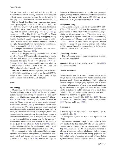
(Tanaka et al. 2010; Plate 1).<br />
Concluding remarks<br />
The Melanomma concept based on ascospore morphology<br />
appears polyphyletic.<br />
Metameris Theiss. & Syd., Annls mycol. 13: 342 (1915).<br />
(Phaeosphaeriaceae)<br />
Generic description<br />
Habitat terrestrial, saprobic or parasitic. Ascostromata erumpent<br />
through the host surface in linear rows parallel to the host fibers.<br />
Ascomata small, globose to subglobose, black, coriaceous.<br />
Peridium composed of large lightly pigmented cells of textura<br />
angularis. Hamathecium of rare, broad pseudoparaphyses,<br />
septate, constricted at the septa. Asci bitunicate, fissitunicate,<br />
broadly cylindrical to slightly obclavate, with a short, thick,<br />
knob-like pedicel. Ascospores hyaline, 1- (rarely 2-) septate.<br />
Anamorphs reported for genus: none.<br />
Literature: von Arx and Müller 1975; Barr1972; Clements<br />
and Shear 1931; Eriksson 2006; Lumbsch and Huhndorf<br />
2007; TheissenandSydow1915.<br />
Type species<br />
Metameris japonica (Syd.) Syd., Annls mycol., 13(3–4):<br />
342 (1915). (Fig. 59)<br />
≡ Monographus japonicus Syd. Annls mycol. 10: 408<br />
(1912).<br />
Ascostromata erumpent through the host surface in linear<br />
rows parallel to the host fibers, 500–750 μm long and 140–<br />
200 μm wide, with three to ten ascomata arranged in a line<br />
(Fig. 59a). Ascomata 115–160 μm diam., semi-immersed in<br />
substrate to erumpent, globose, subglobose, black, coriaceous<br />
(Fig. 59b). Cells of ascostromata heavily pigmented and<br />
thick-walled, cells of peridium composed of large lightly<br />
pigmented cells of textura angularis, cells 5–15 μm diam.,


